
|
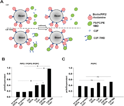
Fig. 7 Reconstituted otoferlin docks adjacent liposomes. (A) Diagram of the pull-down system. Rhodamine-labeled liposomes are conjugated to streptavidin beads and mixed with either NBD liposomes and soluble C2F (top) or C2F-TMD proteoliposomes labeled with NBD (bottom). (B) Quantitation of liposome sedimentation assay with bead-conjugated liposomes with PI(4,5)P2 (5:25:60:5:5 PIP2, POPS, POPC, rhodamine-PE, biotin-PE) (t-test, p < 0.001). (C) Quantitation of liposome sedimentation assay with bead-conjugated liposomes without negatively charged lipids (90:5:5 POPC, rhodamine-PE, biotin-PE) (t-test, p < 0.001). N = 4.