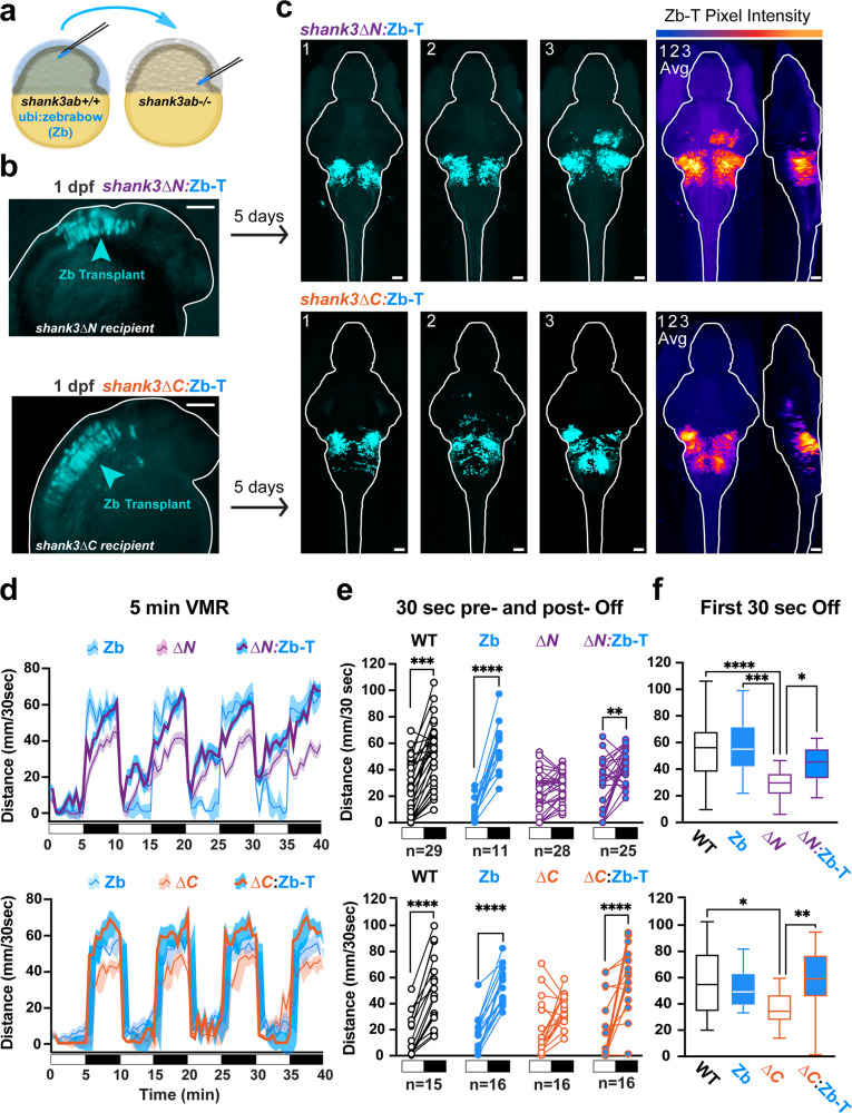
Fig. 3 a A cartoon shows how cells from wild type donor embryos marked by a ubiquitously expressed dTomato fluorescent protein (ubi:zebrabow) are transplanted into the presumptive hindbrain of shank3ab−/− mutant recipient embryos at mid-gastrulation stages. b Chimeric embryos at 1 day post-fertilization (dpf), with donor cells expressing the fluorescent protein (false-colored in cyan) in recipient shank3abΔN−/− or shank3abΔC−/− embryos. Chimeric six-day-old larvae (shank3ab−/−:Zb-T) were imaged to determine the fate of the transplanted cells. c Confocal images of chimeric larvae at 6 dpf following behavioral screening, demonstrating transplanted cells in rescued larvae populate the dorsal/rostral brainstem nuclei. Individual representative larvae are numbered 1-3, with the three averaged in the right most stack. d VMR line graphs, median +/− SE, (d–f) and (e) paired dot plots show lights-off behavioral phenotypes are rescued in both shank3abΔ−/− mutant models with wild-type-derived brain stems (shank3abΔ−/−:Zb-T). Exact sample sizes of biologically independent samples for each genotype and chimera are indicated below each plot and also apply to d and f. Within shank3 model comparisons were conducted using Dunn–Bonferroni p-value corrected t-tests. f Box plots displaying median swimming distances for individuals following the first 30 s following lights-off. Individual values are medians representing all four lights-off transitions for individual larvae. Boxes denote the median, 1st and 3rd quartile, while whiskers represent the minimum and maximum values. Groups were statistically compared using Kruskal-Wallis one-way ANOVA, and when p < 0.05, were followed by Dunn’s multiple comparisons. P-value asterisks represent; p < 0.05 - *, p < 0.01 - **, p < 0.001 - ***, p < 0.0001-****. Scale bars = 100 µm (b); 50 µm (c). Source data for plots are provided in Supplementary Data 2.
Image
Figure Caption
Acknowledgments
This image is the copyrighted work of the attributed author or publisher, and
ZFIN has permission only to display this image to its users.
Additional permissions should be obtained from the applicable author or publisher of the image.
Full text @ Commun Biol

