
|
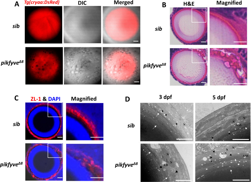
Figure 4.
(A) Confocal imaging of the lens of 5-dpf siblings and pikfyveΔ8 mutants in Tg(cryaa:DsRed) transgenic background. (B) Hematoxylin-eosin (HE) staining of 5-dpf siblings and pikfyveΔ8 mutant zebrafish lens after cryostat section. (C) ZL-1 antibody and DAPI staining of 5-dpf siblings and pikfyveΔ8 mutant zebrafish lens. (D) Transmission electron microscope (TEM) images of the lens of siblings and pikfyveΔ8 mutants at 3 dpf and 5 dpf. ASS, autophagy lysosome; LD, lipid droplet; N, nucleus. All results were confirmed in three different individuals. All the scale bars represent 10 μm.Detailed characterization of cataract phenotypes in pikfyveΔ8 mutants.