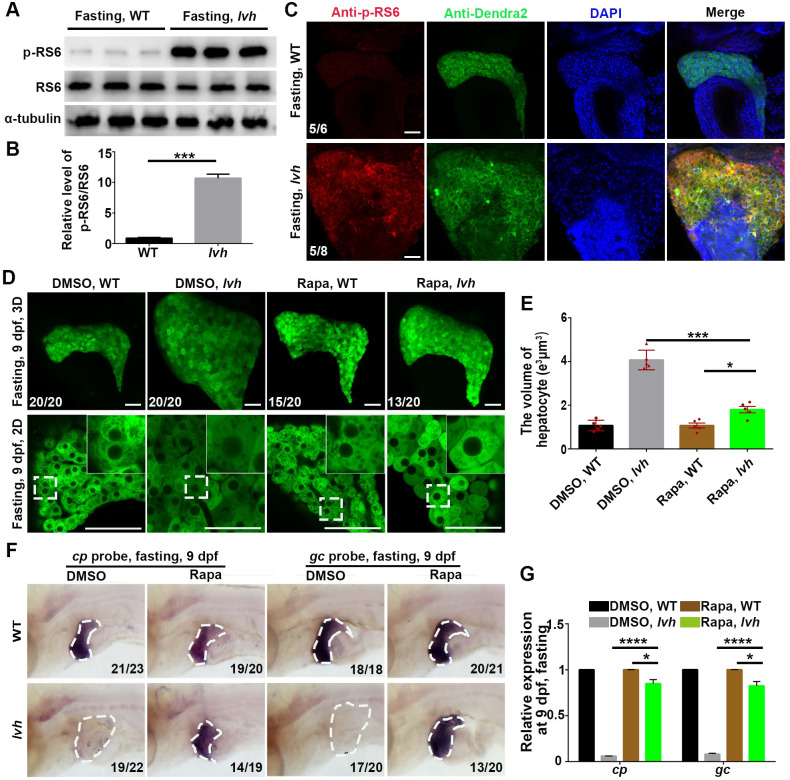
Fig 4 FTCD prevents starvation-induced hepatomegaly through downregulating mTORC1.(A) Western blot analysis of p-RS6 (s240/244), RS6, and α-tubulin using liver lysates (n = 120). (B) Quantification of relative intensity (n = 3) for p-RS6/RS6. (C) Immunostaining for p-RS6 and Dendra2 in livers (3D imaging). Nuclei were stained with DAPI (blue). (D) 3D confocal projection and 2D single-optical section images of the liver. Higher magnification images of single hepatocytes are displayed. (E) Unpaired Student’s t-test for single hepatocyte volume of DMSO and Rapa treatment in the wild-type (n = 5) and lvh (n = 5). (F) The disappeared cp and gc expressions in lvh under fasting were rescued by rapamycin treatment. The dashed boxes indicate the liver area. (G) qPCR data showing the relative expression levels of cp and gc in the wild-type and lvh liver after DMSO and Rapa treatment. Asterisks indicate statistical significance. NS, not significant. *P<0.05, ***P<0.001, ****P<0.0001. Data are represented as mean±SD. WT, wild-type. Rapa, rapamycin. Scale bars, 50 μm
Image
Figure Caption
Figure Data
Acknowledgments
This image is the copyrighted work of the attributed author or publisher, and
ZFIN has permission only to display this image to its users.
Additional permissions should be obtained from the applicable author or publisher of the image.
Full text @ PLoS Genet.

