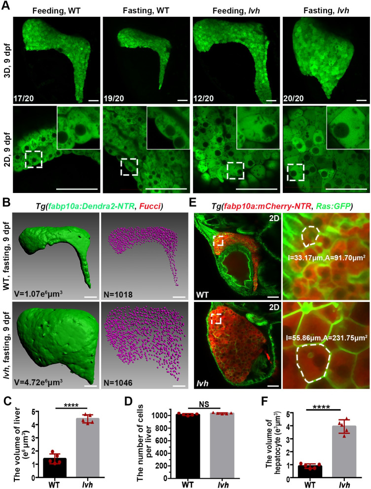
Fig 1 The lvh mutant exhibits hepatomegaly and enlarged hepatocyte size under fasting. (A) Confocal 3D projection and 2D single-optical section images of the liver under feeding and fasting in the wild-type or lvh mutant at 9 dpf. Higher magnification images showing single hepatocytes are displayed upright. (B) 3D reconstruction images showing the liver volumes and hepatocyte nuclei of the wild-type and lvh mutant at 9 dpf. (C) Unpaired Student’s t-test for the liver volume of wild-type (n = 5) and lvh (n = 5). (D) Unpaired Student’s t-test for the number of hepatocytes per liver of wild-type (n = 5) and lvh (n = 5). (E) Single-optical section images showing livers of the wild-type and lvh mutant at 9 dpf. Higher magnification images of single hepatocytes (dashed frames) are displayed. (F) Unpaired Student’s t-test for single hepatocyte volume in the wild-type (n = 5) and lvh (n = 5). NS, not significant. ****P<0.0001. WT, wild-type. Data are represented as mean±SD. Scale bars, 50 μm.
Image
Figure Caption
Figure Data
Acknowledgments
This image is the copyrighted work of the attributed author or publisher, and
ZFIN has permission only to display this image to its users.
Additional permissions should be obtained from the applicable author or publisher of the image.
Full text @ PLoS Genet.

