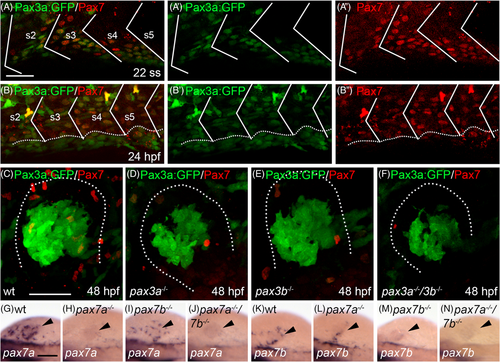
Fig. 6 Pax7 and Pax3a:GFP are co-expressed in the somite before migrating muscle progenitors (MMPs) start to migrate out. Lateral view of somites 2-5 showing transgenic expression of A Pax3a:GFP (green) and immunohistochemical detection of Pax7 (red), A', Pax3a:GFP and, A", Pax7 in zebrafish embryos at the 22 somite stage (ss). Lateral view of somites 2-5 showing transgenic expression of B Pax3a:GFP (green) and immunohistochemical detection of Pax7 (red), B', Pax3a:GFP and, B'', Pax7 in zebrafish embryos at 24 hpf. Myosepta are marked with white lines and dashed line in B indicates yolk-somite border. Transgenic expression of Pax3a:GFP (green) and immunohistochemical detection of Pax7 (red) in C wt, D, pax3a−/−, E, pax3b−/− and, F, pax3a−/−/3b−/− double mutant pectoral fin bud at 48 hpf. Dotted line in C-F indicates outline of fin bud. Expression of pax7a in, G, wt (n = 65), H, pax7a−/− (n = 8), I, pax7b−/− (n = 18) and, J, pax7a−/−/7b−/− (n = 4). Expression of pax7b in (K) wt (n = 101), L, pax7a−/− (n = 13), M, pax7b−/− (n = 12), and N pax7a−/−/7b−/− (n = 5). Arrowhead indicates location of fin bud. Scale bar: A-F, 50 μm, G-N, 100 μm
Image
Figure Caption
Figure Data
Acknowledgments
This image is the copyrighted work of the attributed author or publisher, and
ZFIN has permission only to display this image to its users.
Additional permissions should be obtained from the applicable author or publisher of the image.
Full text @ Dev. Dyn.

