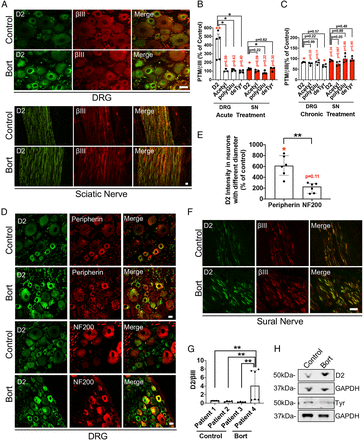
Fig. 1 Bort induces D2 levels in vivo. (A) Representative D2 and βIII IF staining of DRG cell bodies and the SN isolated from control rats or rats acutely treated with Bort (i.v. 0.2 mg/kg; 24 h). (B and C) Relative tubulin PTM levels measured by quantitative IF in randomly selected DRG cell bodies (35–60) and SN (five sections per condition) from rats treated with acute (n = 3 to 5 per group) (B) (i.v. 0.2 mg/kg; 24 h) or chronic (n = 4 per group) (C) (0.2 mg/kg, 3× week for 8 wk) doses of Bort. (D) Representative peripherin (smaller unmyelinated C-fibers), NF200 (medium and large myelinated A-β fibers), and D2 IF staining of DRG cell bodies acutely treated with Bort. (E) Relative D2 tubulin values measured in DRG cell bodies (n = 35 to 60) positive to either peripherin or NF200 from rats (n = 6) acutely treated with Bort (i.v. 0.2 mg/kg; 24 h). (F) Representative D2 and βIII IF staining of sural nerve biopsy from a patient with BIPN. (G) Ratio analysis of D2/βIII levels measured by IF of fixed tissue (three to six sections per condition) from one sural nerve biopsy of a BIPN patient versus three sural nerve biopsies from control patients. (H) Immunoblot analyses of D2 levels in whole cell lysates from one sural nerve biopsy from a patient treated with Bort displayed with a control patient. Tyr, tyrosinated tubulin. GADPH, loading control. All data in B, C, E, and G are shown as medians plus interquartile range, and statistics were analyzed by Mann–Whitney U test. *P < 0.05, **P < 0.01. Scale bars in A, D, and F, 20 μm. Red writings are P values when compared to control; black writings are P values when compared between groups.
Image
Figure Caption
Acknowledgments
This image is the copyrighted work of the attributed author or publisher, and
ZFIN has permission only to display this image to its users.
Additional permissions should be obtained from the applicable author or publisher of the image.
Full text @ Proc. Natl. Acad. Sci. USA

