Figure 4
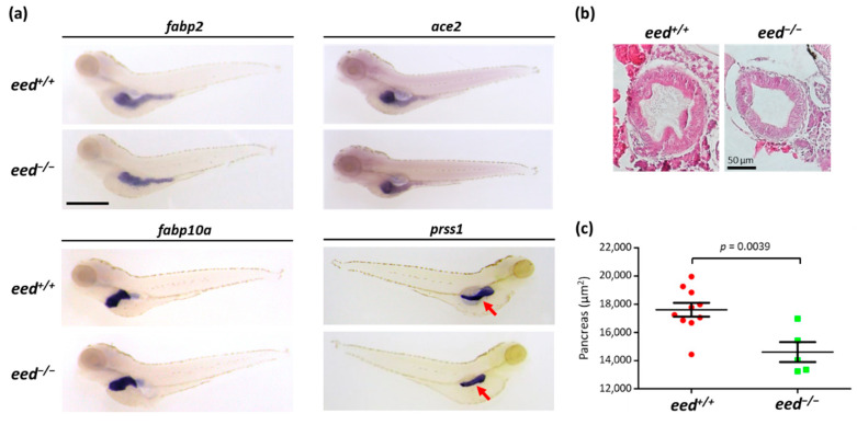
Organization of the digestive organs at 5 dpf: (a) whole-mount RNA in situ hybridization to detect the expression of markers of the intestine (fabp2, ace2), the liver (fapb10a), and the exocrine pancreas (prss1) in eed+/+ and eed−/− siblings at 5 dpf. The red arrow shows the pancreas. Scale bar is 500 µm; (b) intestinal bulb sections from eed+/+ (left) and eed−/− (right) larvae at 5 dpf stained with hematoxylin and eosin. Scale bar is 50 µm; (c) measurement of the surface of the pancreas labeled by in situ hybridization using a prss1 probe at 5 dpf for eed+/+ (red) and eed−/− (green) larvae. Statistical significance was assessed by a Student t-test, and the corresponding p-value is indicated.

