Image
Figure Caption
Fig. 2.
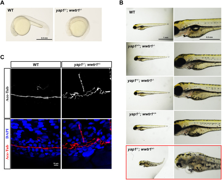
Functional redundance of yap1 and wwtr1 in pronephros development at embryonic stage. (A) Phenotype analysis of yap1−/−;wwtr1−/− embryos at 20 hpf. The embryos of yap1−/−;wwtr1−/− double mutant exhibited no tail extension. Scale bar: 0.5 mm. (B) Phenotype analysis of embryos with different genotypes produced by yap1+/−;wwtr1+/− in-cross. Scale bars: 1 mm (left) and 0.5 mm (right). (C) Whole-mount immunofluorescent staining of acetylated tubulin (red) and DAPI (blue) in yap1−/−;wwtr1+/− embryo and WT embryo at 48 hpf. Z projection of 0.5 μm optical section stacks. Scale bar: 10 μm.
Acknowledgments
This image is the copyrighted work of the attributed author or publisher, and
ZFIN has permission only to display this image to its users.
Additional permissions should be obtained from the applicable author or publisher of the image.
Full text @ Dis. Model. Mech.

