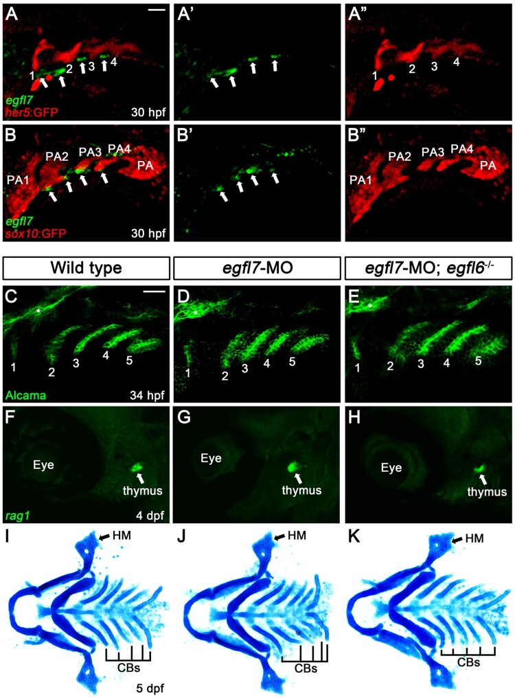
Figure 4.
Expression of egfl7 in the pharyngeal region. (A, B) Fluorescence in situ hybridization of egfl7 (green) in conjunction with the GFP immunohistochemistry (red) in wild-type animals at 30 hpf. (A) egfl7 is expressed segmentally in small patches (arrows) adjacent to her5-positive pouches (1-4). (B) egfl7 expressing small patches (arrows) are located at the ventral tip of sox10-positive pharyngeal arches (PA2-4) but rarely overlapped with PAs. (C-E) Alcama immunohistochemistry (green) labels five pouches (1-5) in wild-type (n = 92), egfl7-MO (n = 80), and egfl7-MO-injected egfl6 mutant (n = 21) embryos at 34 hpf. Sensory ganglia are indicated with asterisks. (F-H) At 4 dpf, rag1 expression (green) in the thymus is normal in wild-type (n = 74), egfl7-MO (n = 76), and egfl7-MO-injected egfl6 mutant (n = 14) zebrafish. (I-K) Facial cartilages, including the HM and CBs, are normal in wild-type (n = 105), egfl7-MO (n = 84), and egfl7-MO-injected egfl6 mutant (n = 19) animals at 5 dpf. Scale bar: 40 μm.

