
|
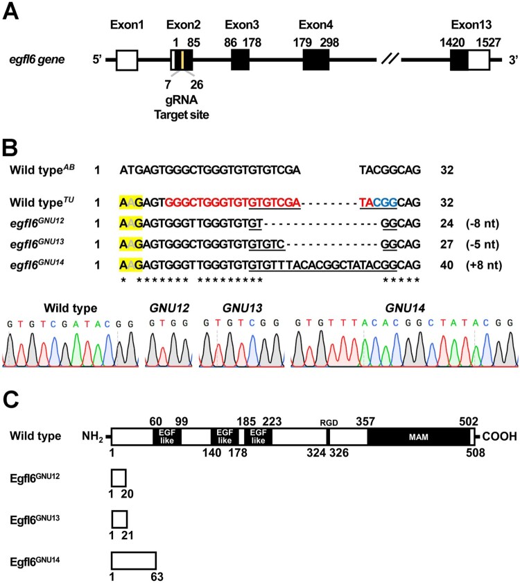
Figure 2.
Generation of loss-of-function mutations in egfl6 gene. (A) Structure of egfl6 gene. egfl6 gene consists of 13 exons bearing sequences for the protein-coding region (black box) and the 5′ and 3′ untranslated regions (open box). The gRNA target site in the second exon is marked in yellow. (B) Mutant alleles of egfl6 gene. The in/del mutation of each mutant allele is shown in the multiple sequence alignments, with the gRNA target and the PAM sites being marked in red and blue, respectively in the wild-typeTU egfl6 sequence. The electrophoretograms show the lesion in each egfl6 mutant allele that is underlined in the multiple sequence alignments. The start loss mutation in egfl6 gene in the wild-type TU strain is highlighted in yellow. (C) Schematic of the Egfl6 protein encoded by the wild-type and mutant alleles. The conserved five domains marked in the wild-type Egfl6 protein are missing in all mutant Egfl6 proteins.