IMAGE

Figure 1

ID
ZDB-IMAGE-210818-37
Source
Figures for Seese et al., 2021
Image
Figure Caption

Figure 1

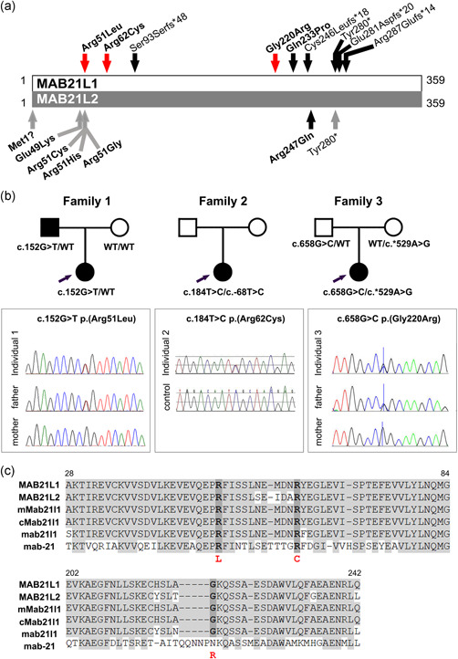
MAB21L1variant details. (a) Schematic of MAB21L1 (top) and MAB21L2 (bottom) proteins. Newly identified heterozygous MAB21L1 coding variants are indicated by a red arrow; previously reported MAB21L1 and MAB21L2 recessive variants are indicated with a black arrow; previously reported MAB21L2 dominant variants are indicated with a gray arrow. Missense variants are bolded. (b) Pedigrees for Families 1–3 indicating MAB21L1 genotype and Sanger sequencing traces for the identified coding variants. (c) Alignment of MAB21L1 and related proteins showing conservation at and around the Arg51Leu, Arg62Cys, and Gly220Arg variants. Identical amino acids are shaded in gray; positions of variant amino acids are indicated with dark gray. Human MAB21L1 (NP_005575.1), human MAB21L2 (NP_006430.1), mouse Mab21l1 (NP_034880.1), chicken Mab21l1 (NP_989864.1), zebrafish mab21l1 (NP_694506.2), and Caenorhabditis elegans mab‐21 (NP_497940.2) are shown

Acknowledgments
This image is the copyrighted work of the attributed author or publisher, and ZFIN has permission only to display this image to its users. Additional permissions should be obtained from the applicable author or publisher of the image. Full text @ Hum. Mutat.