Figure Caption
Fig. 1.
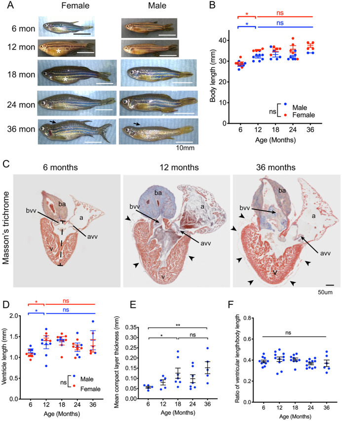
Zebrafish do not exhibit indeterminate somatic or ventricular growth. (A) External appearances of male and female zebrafish between 6 months and 36 months of age. Female fish have larger abdomen than male fish (asterisk). Spinal deformities are more common and increasingly severe after 24 months (arrows). (B) Lengths of male (blue points) and female (red points) zebrafish with increasing age. Sample size (n) is 11, 11, 10, 11, 6 for age groups 6, 12, 18, 24 and 36 months, respectively. There were no differences between the lengths of male and female fish at any stage. After a rapid increase in length in the first 6 months, there was a smaller increase in length in the second 6 months of life, but no change thereafter. (C) Midline histological sections through zebrafish hearts stained with Masson's trichrome. Myocardium is indicated in brown and collagen in blue. The 6-month-old heart had minimal collagen staining in the bulbus arteriosus (ba). Epicardial adipose tissue is not seen. At 12 months, the heart was larger and the bulbus stained strongly for collagen fibres, and there is some epicardial adipose tissue (arrowheads). In the 36-month-old heart, there were extensive epicardial adipose deposits seen on the surface of the heart (arrowheads). There were also extensive collagen deposits in the bulbus arteriosus (ba), bulbo-ventricular valve (bvv) and atrioventricular valve (avv). (D) Ventricular length measured from the histological sections in C, from apex to base of chamber, indicated by the dashed bar in C (6 months). All individual data points are shown (male, blue points; female, red points). Sample size (n) was 11, 11, 10, 11 and 6 for age groups 6, 12, 18, 24 and 36 months, respectively. Ventricular length increased between 6 and 12 months but not thereafter, and there was no difference between hearts from male and female fish. (E) Mean thickness of the ventricular myocardium compact layer increased until 18 months of age and did not change thereafter. Sample size (n) was 5, 5, 7, 7 and 5 for age groups 6, 12, 18, 24 and 36 months, respectively. (F) Ratio of ventricular length (D) to body length (B). Sample size (n) is 10, 11, 9, 11 and 6 for age groups 6, 12, 18, 24 and 36 months, respectively. There was no difference in the ratio throughout adult life. All individual data points are shown. Data are mean±s.e.m. *P<0.05; **P< 0.02; ns, not significant (two-way ANOVA was used in B,D; one-way ANOVA in E,F). a, atrium; avv, atrioventricular valve; ba, bulbus arteriosus; bvv, bulbo-ventricular valve; v, ventricle.
Acknowledgments
This image is the copyrighted work of the attributed author or publisher, and
ZFIN has permission only to display this image to its users.
Additional permissions should be obtained from the applicable author or publisher of the image.
Full text @ Dis. Model. Mech.