
|
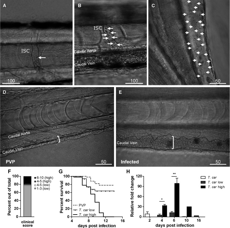
Figure 2—figure supplement 1 Tg(mpeg1:mCherryF;mpx:GFP) were injected intravenously at 5 dpf with n = 200 T. carassii or with PVP. At 4 dpi, larvae were separated in high- and low-infected individuals based on our clinical scoring criteria. At each time point, three pools of 3–5 larvae were sampled for subsequent real-time quantitative gene expression analysis. Each data point represents the mean of three pools, except for the high-infected group at 10 dpi where only one pool could be made due to low survival. Relative fold change was normalised relative to the zebrafish-specific ef1α housekeeping gene and expressed relative to the PVP-injected group at each time point.