IMAGE

Figure 2 supplement 1

ID
ZDB-IMAGE-210625-21
Genes
Source
Figures for Gentile et al., 2021
Image
Figure Caption

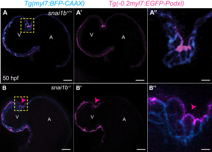
Figure 2 supplement 1 Apicobasal polarity is maintained in extruding cardiomyocytes (CMs) in <italic toggle='yes'>snai1b</italic> mutants.

(A–B’’) Single-plane images of Tg(myl7:BFP-CAAX); Tg(−0.2myl7:EGFP-podocalyxin) snai1b+/+ (A–A’) and snai1b-/- (B–B’) hearts at 50 hpf. Close-up of boxed areas (A’’, B’’). No changes in the localization of the apical marker Podocalyxin are observed in extruding CMs (arrowheads in B–B’’). Scale bars: 20 µm (A–A’, B–B’), 2 µm (A’’, B’’). V: ventricle; A: atrium.

Figure Data
Acknowledgments
This image is the copyrighted work of the attributed author or publisher, and ZFIN has permission only to display this image to its users. Additional permissions should be obtained from the applicable author or publisher of the image. Full text @ Elife