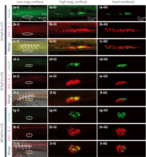
Fig. 1 Morphology of the pancreatic islet and β-cells at 24, 30 and 48 h post-fertilization (hpf). Embryos were fixed at (a–c) 24 hpf (n = 37), (d–f) 30 hpf (n = 55) and (g–i) 48 hpf (n = 23). They were then de-yolked and whole-mount double-immunolabeled with antibodies against insulin (green) and islet 1 and 2 (red) to show the localization of the β-cells and islet cells in the endocrine pancreas, respectively. All images were acquired with the Leica TCS SP5 II laser scanning confocal system. The images shown in panels a-i to i-i were acquired at low magnification (i.e., with a ×20 HC PL APO objective lens), whereas those shown in panels a-ii to i-ii were acquired at a higher magnification (i.e., with a ×63 HCX PL APO water-dipping objective lens). These are all single optical sections taken through the middle of the pancreatic islet. The images shown in panels a-iii to i-iii are stacks of optical images rendered as 3-D images by the image analysis software, Imaris. (a,d,g) The localization of insulin-positive β-cells (in green). (b,e,h) The localization of the islet cells (in red). (c-i,f-i,i-i) The co-localization of insulin, islet 1 and 2 and bright-field image. (c-ii,f-ii,i-ii) The co-localization of insulin and islet 1 and 2. D, dorsal; Ant., anterior; Pos., posterior; S, somite; V, ventral.
Image
Figure Caption
Figure Data
Acknowledgments
This image is the copyrighted work of the attributed author or publisher, and
ZFIN has permission only to display this image to its users.
Additional permissions should be obtained from the applicable author or publisher of the image.
Full text @ J Diabetes Investig

