
|
Fig. 3
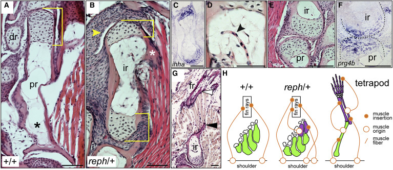
Figure 3. Intermediate radials are long bones morphologically integrated into the fin (A–G) Histology of adult fins. (A) Wild-type proximal radial with a single distal epiphysis (bracket). (B) reph/+ intermediate radial with dual epiphyses (brackets), a muscle insertion point (asterisk), and distal synovial cavity (arrowhead). (C) ihha expression in epiphyses of an intermediate radial. (D) Blood vessel invading an intermediate radial (black arrowhead). (E) Synovial joint between intermediate and proximal radials. (F) prg4b expression in the proximal-intermediate radial joint. (G) Muscle fibers originating from process on intermediate radial (black arrowhead) and inserting on the dermal fin rays (white arrowhead). (H) Diagram of muscle attachment in the tetrapod limb versus wild-type and reph/+ zebrafish pectoral fins. Anterior to left, distal to top; dr, distal radial; fr, fin ray; ir, intermediate radial; pr, proximal radial; scale bars 50 μm.
Reprinted from Cell, 184(4), Hawkins, M.B., Henke, K., Harris, M.P., Latent developmental potential to form limb-like skeletal structures in zebrafish, 899-911.e13, Copyright (2021) with permission from Elsevier. Full text @ Cell