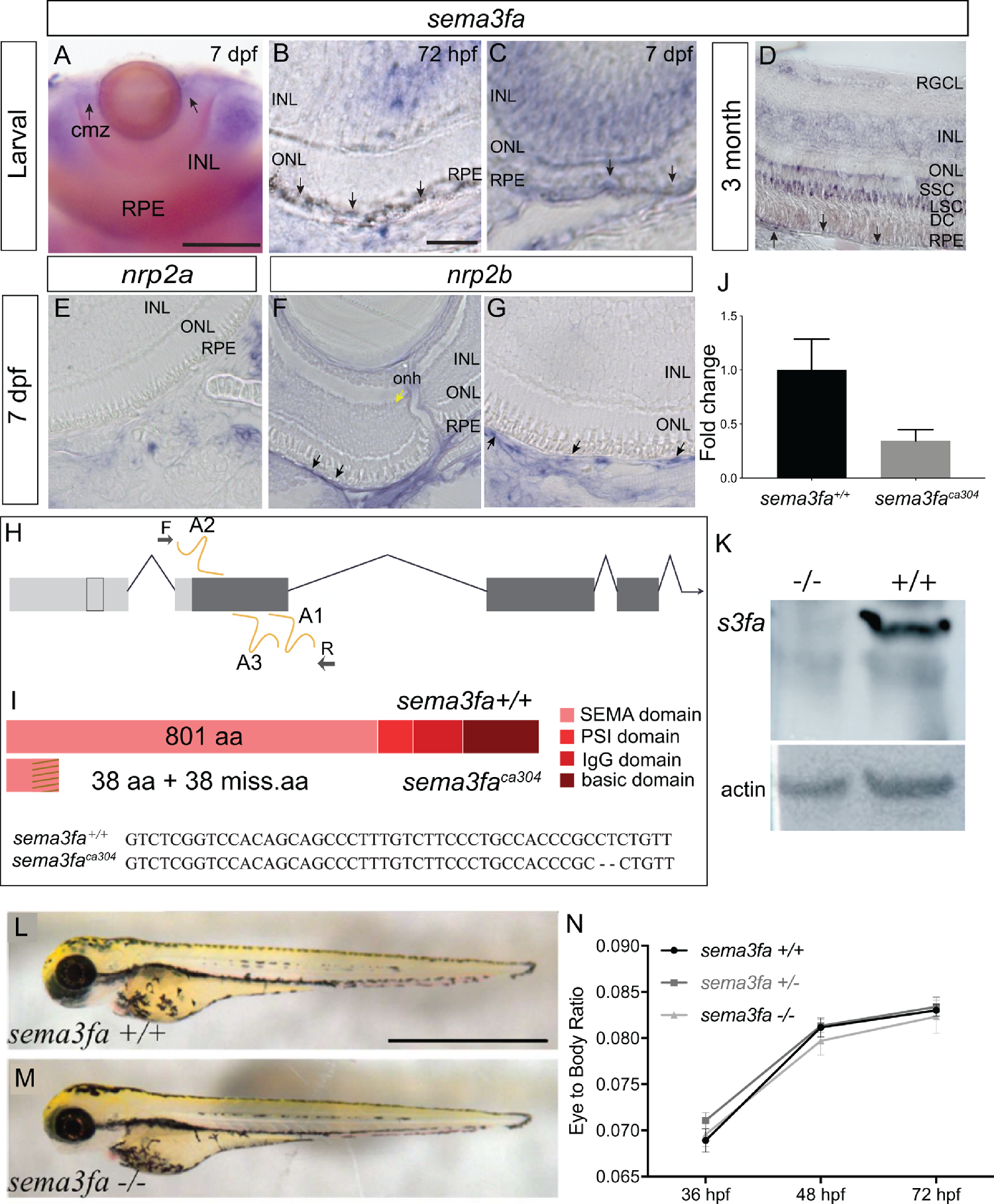
Fig. 1 Sema3fa is expressed by the RPE and inner retina of larval and adult zebrafish. (A–D) sema3fa RNA ISH in whole mount (A) and retinal sections (B–D). ISH signal is detected in the retinal pigment epithelium (RPE) during development (A–C) and into adulthood (D). In larval and adult retina, sema3fa is also expressed strongly in the inner nuclear layer (INL) and CMZ, with expression in the outer nuclear layer (ONL) observed in the adult. (E–G) Expression at 7 dpf of mRNA for the known Sema3fa receptor nrp2b (F,G) but not nrp2a (E) by the endothelial cells of the retinal artery that runs alongside the optic nerve head (yellow arrow) and the choroid vessels lining the back of the eye (black arrows). Note that to minimize the pigment of the RPE, retinal sections were either from larvae treated at 24 hpf with 1-phenyl-2-thiourea (A–C, E–G), or were bleached (D). (H) Chromosomal overview of the sema3fa locus targeted by CRISPR/Cas9 mutagenesis to exon 1 (guide RNA: A1-3) and primers used to identify the mutation. UTR: untranslated region; F: forward primer; R: reverse primer. (I) Schematic representation of WT and premature stop codon mutant proteins. sema3faca304 fish have a 2 bp deletion (dashes) and produce a predicted product of 76 aa. Dashes represent missense amino acids (miss.aa). (J) RT-qPCR of sema3fa mRNA levels in WT and sema3fbca304 embryos at 48 hpf suggest nonsense mediated decay of mRNA transcript (N = 3). Error bar represents standard error of the mean (SEM). (K) Western blot of protein isolated from 5-6 dpf WT and sema3faca304 mutant fish processed with a custom-made antibody against zebrafish Sema3fa. The antibody recognizes a protein of the appropriate size for Sema3fa in WT fish, which is absent in the mutants. Loading control is ß-actin. (L, M) Lateral views of 72 hpf WT (H) and sema3faca304 (I) fish. (N) The ratio of eye width to anterior-posterior body length, measured along the antero-posterior axis, at 36, 48, and 72 hpf, arguing that there are no gross abnormalities in body or eye growth in the sema3fa heterozygote (n = 10) and homozygote (n = 4) mutant embryos as compared to WT (n = 10). Scale bars: A: 100 µm; B: 15 µm; L: 1 mm.
Image
Figure Caption
Figure Data
Acknowledgments
This image is the copyrighted work of the attributed author or publisher, and
ZFIN has permission only to display this image to its users.
Additional permissions should be obtained from the applicable author or publisher of the image.
Full text @ Invest. Ophthalmol. Vis. Sci.

