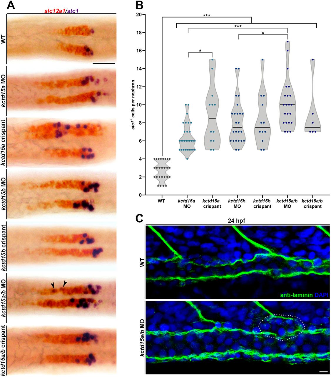
Fig. 3 kctd15a/b deficiency elevates CS differentiation. (A) Whole-mount in situ hybridization of slc12a1 (red) and stc1 (purple) at 24 hpf in pronephric cells of wild type, three kctd15 MO and three kctd15 crispants. Black arrowheads indicate proximal stray stc1+ cells clearly separate from the main CS cluster in kctd15a/b MO. Scale bar: 35 μm. (B) Quantification of stc1+ cell number per nephron. Black brackets cluster groups together for collective comparison. (C) Immunofluorescence of laminin (green) in the pronephros of 24 hpf wild type and kctd15a/b MO. Premature basement membrane formation occurs in the kctd15a/b morphant, which separates the budding CS cell cluster (white dotted circle) from the pronephric tubule. Scale bar: 5 μm. n≥3. *P<0.05; ***P<0.001. Data are mean±s.d. Cell counts were compared using ANOVA. Data are displayed as individual points distributed about the mean (black line).
Image
Figure Caption
Acknowledgments
This image is the copyrighted work of the attributed author or publisher, and
ZFIN has permission only to display this image to its users.
Additional permissions should be obtained from the applicable author or publisher of the image.
Full text @ Development

