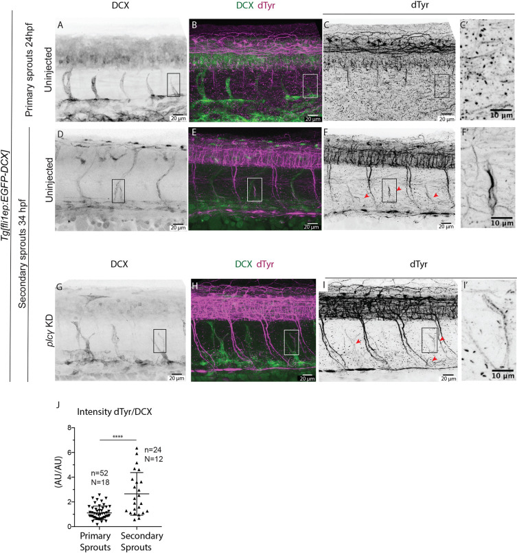
Fig. 5. Microtubules of secondary sprouts are selectively detyrosinated. (A-I′) Immunostainings using antibody detecting the glutamate amino acid of detyrosinated microtubules (dTyr) during primary (A-C) and secondary (D-I) sprouting in uninjected (A-F) and plcγ KD (G-I) Tg[fli1ep:EGFP-DCX] embryos, labelling all endothelial microtubules (DCX). C′,F′ and I′ are magnifications from boxed region in C,F,I, respectively. plcγ KD embryos (G-I′) show reduced primary sprouting, facilitating the visualization and quantification of dTyr signal specifically in secondary sprouts. Arrowheads indicate secondary sprouts. (J) Quantification of the dTyr/DCX signal intensities in primary sprouts of control MO-injected embryos, and secondary sprouts from plcγ MO-injected embryos. n=52 control primary sprouts from 18 embryos, n=24 plcγ KD secondary sprouts from 12 embryos, from one experiment. ****<0.00001 (Mann–Whitney test). Pictures are representative of 3 replicated experiments.
Image
Figure Caption
Figure Data
Acknowledgments
This image is the copyrighted work of the attributed author or publisher, and
ZFIN has permission only to display this image to its users.
Additional permissions should be obtained from the applicable author or publisher of the image.
Full text @ Development

