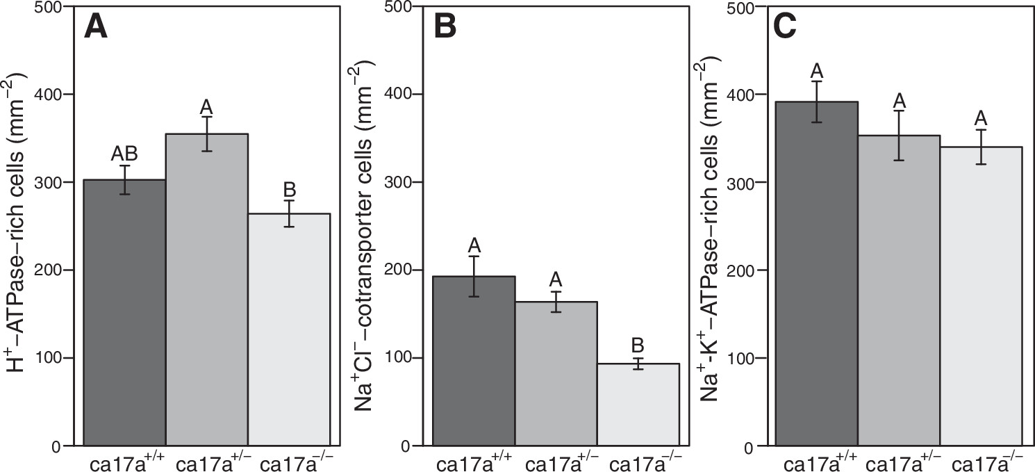
Fig. 6 Ionocyte density in ca17a+/+, ca17a+/−, and ca17a−/− zebrafish (Danio rerio) larvae at 4 dpf. Density of H+-ATPase-rich cells (A) and Na+ Cl−-cotransporter cells (B) significantly differed among genotypes (ANOVA; A: F = 7.7, P < 0.01, n = 14 for ca17a+/+, n = 19 for ca17a+/−, and n = 23 for ca17a−/−; B: F = 20.5, P < 0.01, n = 15 for ca17a+/+, n = 16 for ca17a+/−, and n = 26 for ca17a−/−, respectively). There was no effect of genotype on Na+-K+-ATPase-rich cells (C) (ANOVA; F = 1.3, P = 0.29, n = 8 for ca17a+/+ and ca17a+/−, n = 11 for ca17a−/−). Bars with different letters are significantly different (P < 0.05) from one another across genotypes. Data are presented as means ± SE.
Image
Figure Caption
Figure Data
Acknowledgments
This image is the copyrighted work of the attributed author or publisher, and
ZFIN has permission only to display this image to its users.
Additional permissions should be obtained from the applicable author or publisher of the image.
Full text @ Am. J. Physiol. Regul. Integr. Comp. Physiol.

