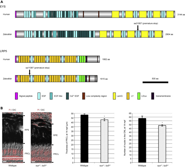
Fig. 1 Figure 1. Conservation of Human and Zebrafish EYS and LRP5 Proteins and Mild Photoreceptor Degeneration in the Digenic eys+/-; lrp5+/− Zebrafish (A) The domain structures of human and zebrafish EYS and LRP5 proteins show that the compositions of EYS and LRP5 proteins are well conserved between the two species. Human and zebrafish EYS proteins consist of a signal peptide sequence for secretion, epidermal growth factor (EGF), EGF-like and Ca2+ EGF domains in the N-terminal half, and laminin A G-like (LamG) and EGF domains in the C-terminal half. Human and zebrafish LRP5 proteins consist of a signal peptide sequence, low-density lipoprotein receptor YWTD (LY; also known as β-propeller), EGF, and LDLa domains in the N-terminal extracellular half followed by transmembrane region and low complexity region in the C-terminal intracellular region. Vertical arrows in zebrafish EYS and LRP5 proteins indicate the point mutations resulting in premature stop codons (nonsense mutations). (B) The thickness of photoreceptor cell layer (PRCL) was 10% reduced in the eys+/-; lrp5+/− zebrafish eyes compared with wild-type PRCL at 14 mpf (wild-type, 53.3 ± 1.1 μm; eys+/-; lrp5+/− mutants, 47.9 ± 1.2 μm). The number of nuclei in the ONL was reduced 16% in the eys+/-; lrp5+/− zebrafish eyes compared with wild-type PRCL (wild-type, 58.3 ± 3.1; eys+/-; lrp5+/− mutants, 49 ± 1.0). Data are represented as mean ± SD. ∗p < 0.01. DIC, differential interference contrast; mpf, months post fertilization; RPE, retinal pigment epithelium. Scale bars, 10 μm.
Image
Figure Caption
Figure Data
Acknowledgments
This image is the copyrighted work of the attributed author or publisher, and
ZFIN has permission only to display this image to its users.
Additional permissions should be obtained from the applicable author or publisher of the image.
Full text @ iScience

