IMAGE

FIGURE 3

ID
ZDB-IMAGE-210108-30
Source
Figures for Zhang et al., 2020
Image
Figure Caption

FIGURE 3

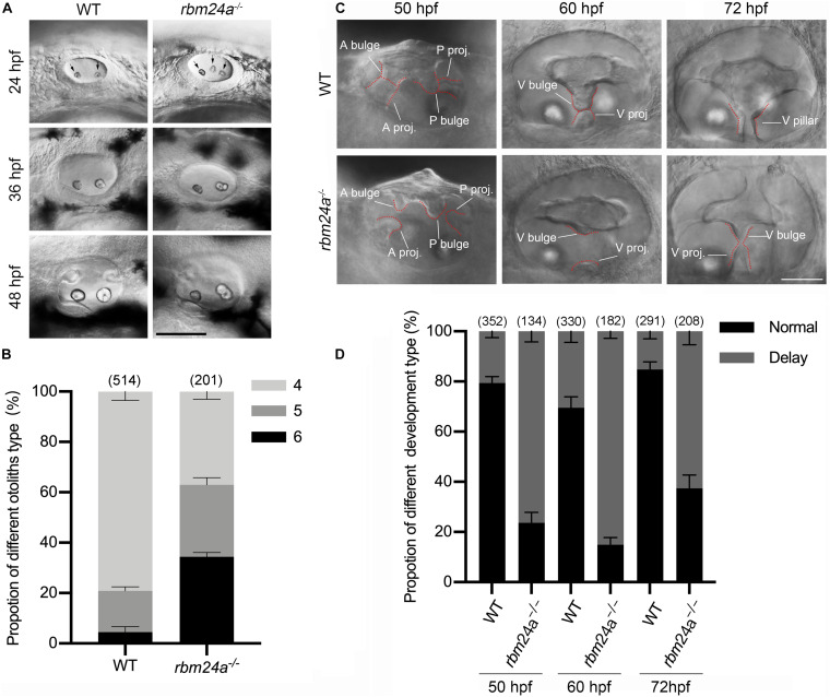
Otolith formation and semicircular canal fusion are delayed in rbm24a mutants. (A) Otolith formation in zebrafish larvae is examined using a light microscope at 24, 36, and 48 hpf as indicated. Otoliths are indicated with arrows. Scale bar, 0.1 mm. (B) The numbers of otic vesicles per larvae at 24 hpf are calculated according to the results from (A). P values are 0.000833 (larvae with 4 otoliths), 0.020678 (larvae with 5 otoliths), and 0.00044 (larvae with 6 otoliths). (C) Fusion of the semicircular canals is examined using a light microscope at 50, 60, and 72 hpf as indicated. Scale bar, 50 μm. (D) The numbers of larvae with normal or delayed semicircular canal fusion are calculated according to the results from (C). P values are 0.000001 (50 hpf), 0.00005 (60 hpf), and 0.000034 (72 hpf). The numbers of larvae for each genotype are indicated in brackets. A bulge, anterior bulge; A proj., anterior projection; P bulge, posterior bulge; P proj., posterior projection; V bulge, ventral bulge; V proj., ventral projection; and V pillar, ventral pillar.

Acknowledgments
This image is the copyrighted work of the attributed author or publisher, and ZFIN has permission only to display this image to its users. Additional permissions should be obtained from the applicable author or publisher of the image. Full text @ Front Cell Dev Biol