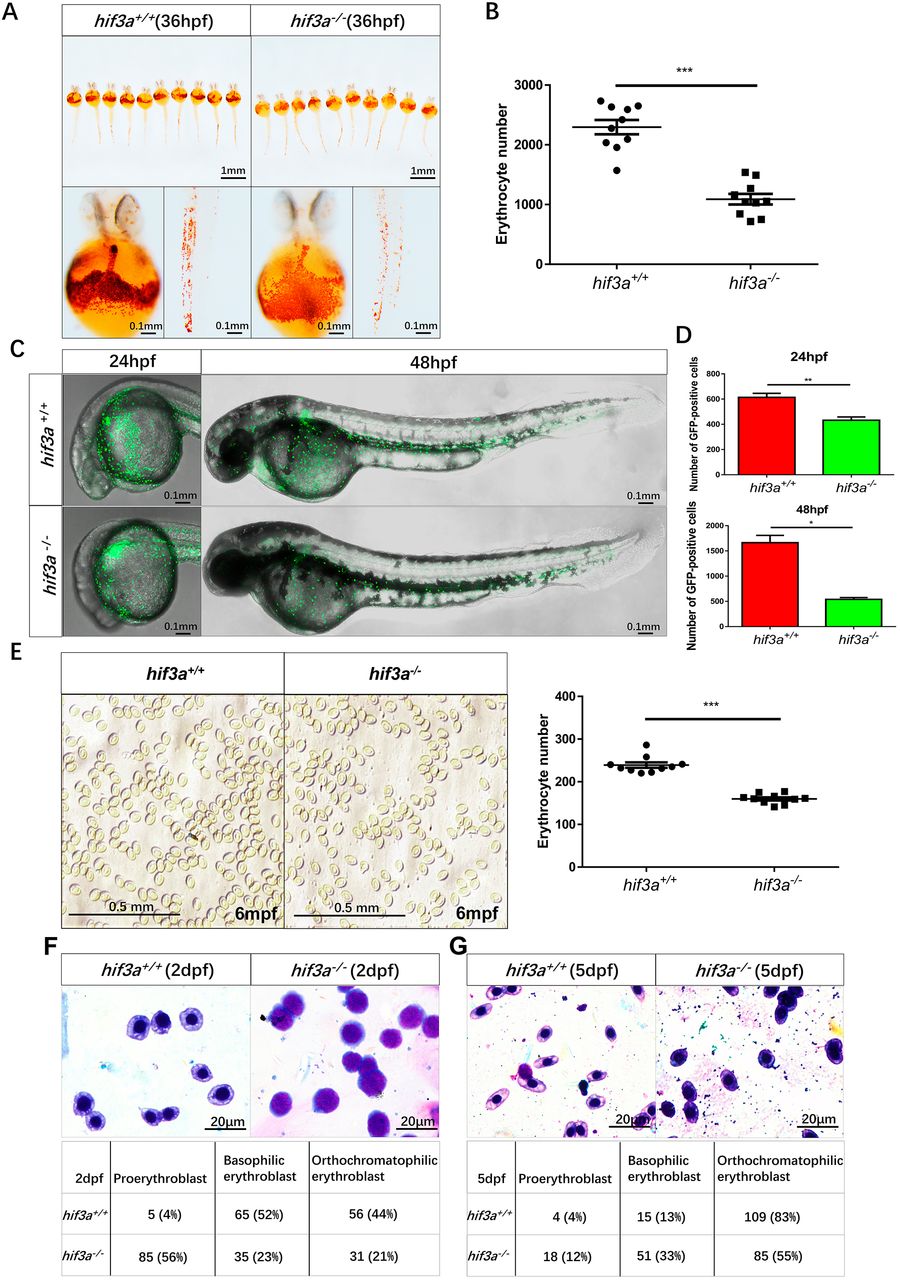
Fig. 2 The number of erythrocytes is reduced and primitive erythroid maturation is retarded in hif3a−/−. (A) O-Dianisidine staining of functional hemoglobin in the mature primitive erythrocytes in hif3a+/+ (left) and hif3a−/− (right) at 36 hpf. The experiments were repeated three times. (B) The number of erythrocytes was reduced in hif3a−/− (10 larvae). (C) Fluorescent images of Tg(gata1:eGFP)/hif3a+/+ (top) and Tg(gata1:eGFP)/hif3a−/− (bottom) indicated that hif3a−/− have fewer gata1-positive erythrocytes at 24 hpf and 48 hpf. (D) Quantitation of gata1-positive erythrocytes in Tg(gata1:eGFP)/hif3a+/+ and Tg(gata1:eGFP)/hif3a−/− at 24 hpf (top) and 48 hpf (bottom) (10 larvae for each, three replicates). (E) The number of erythrocytes was significantly reduced in hif3a−/− compared with hif3a+/+ at 6 mpf. Representative image of erythrocytes in the counting chamber (1 mm×1 mm) (right); scatter plot indicates erythrocyte numbers in 10 counting chambers (left). (F,G) Representative images of May–Grunwald–Giemsa-stained erythroblasts in hif3a+/+ and hif3a−/− larvae at 2 dpf (F) and 5 dpf (G). Cells were morphologically classified at various stages of maturation; numbers (and percentages in brackets) of hif3a+/+ and hif3a−/− larvae at each stage are shown. Error bars indicate the s.e.m.; *P<0.05; **P<0.01; ***P<0.001 (unpaired, two-tailed Student's t-test).
Image
Figure Caption
Figure Data
Acknowledgments
This image is the copyrighted work of the attributed author or publisher, and
ZFIN has permission only to display this image to its users.
Additional permissions should be obtained from the applicable author or publisher of the image.
Full text @ Development

