IMAGE

Figure 3

ID
ZDB-IMAGE-201214-27
Source
Figures for Maradonna et al., 2020
Image
Figure Caption

Figure 3

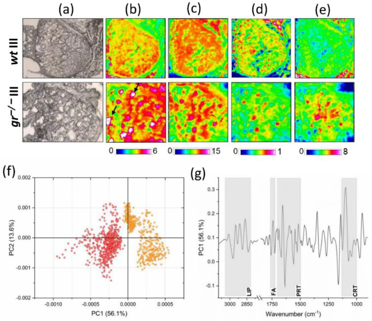
Infrared imaging analysis. (a) Microphotographs of class III oocytes in wt and gr−/− ovary sections. False color images showing the topographical distribution of: (b) lipids (color scale 0–6), (c) proteins (color scale 0–15), (d) glycosylated compounds (color scale 0–1), and (e) cortisol (color scale 0–8) (the dimensions of false color images were 328 × 328 mm). (f) Principal component analysis (PCA) scores’ plot of wt III (orange) and gr−/− (red) spectral data. (g) PC1 loadings of wt and gr−/− class III oocytes.

Acknowledgments
This image is the copyrighted work of the attributed author or publisher, and ZFIN has permission only to display this image to its users. Additional permissions should be obtained from the applicable author or publisher of the image. Full text @ Int. J. Mol. Sci.