
Image
|
Figure Caption
Figure 2
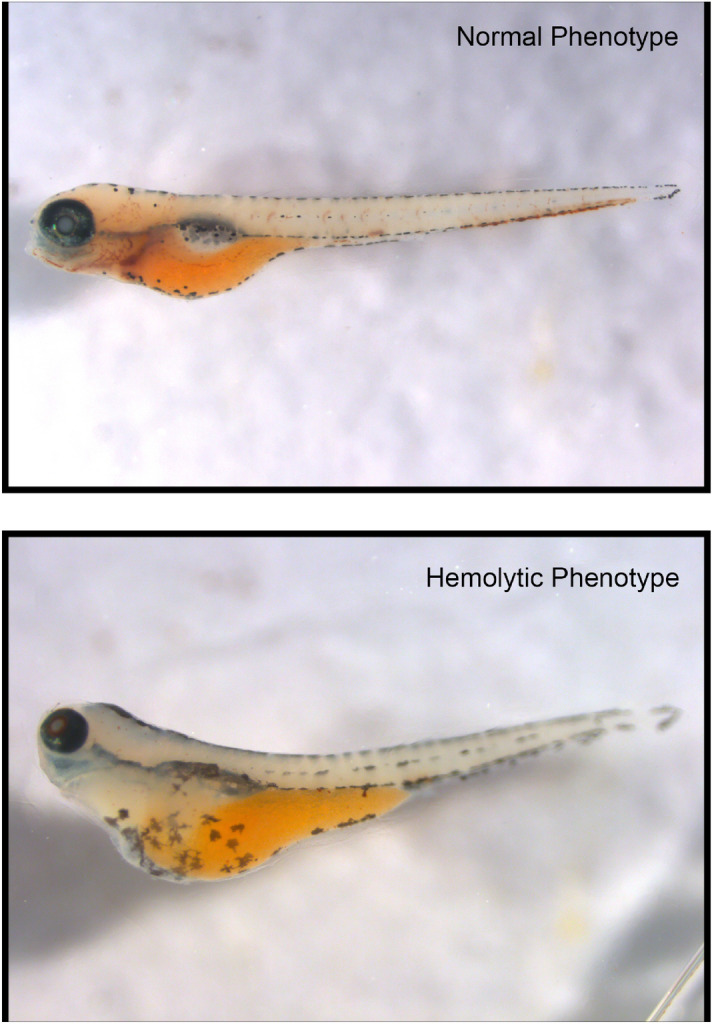
Hemolysis in a G6PD-deficient zebrafish model. Top micrograph shows a zebrafish at 72 hpf with intact red blood cells. Bottom micrograph shows an edematous G6PD-deficient zebrafish at 72 hpf after 48 h of methylated spirit exposure. Both animals were subject to o-dianisidine staining to show hemoglobin containing red cells. Zebrafish were imaged using a Leica DFC340FX fluorescent microscope with PlanAPO 1.6×/0.05 NA objective. Image capture was performed with Leica Application Suite X 3.6.0.2010 (
Figure Data
Acknowledgments
This image is the copyrighted work of the attributed author or publisher, and
ZFIN has permission only to display this image to its users.
Additional permissions should be obtained from the applicable author or publisher of the image.
Full text @ Sci. Rep.