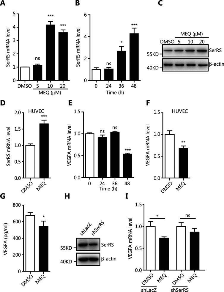
Figure 2
MEQ inhibits VEGFA expression by upregulating SerRS in cultured cells. (A, B) The qRT-PCR shows SerRS expression in MDA-MB-231 cells treated with MEQ at indicated dosages or with dimethyl sulfoxide (DMSO) as a vehicle control for 48 h (A) or treated with 10 μM MEQ for the indicated time periods (B). (C) Western blotting to show SerRS protein levels in MDA-MB-231 cells treated with the indicated dosages of MEQ or DMSO for 48 h. (D) The qRT-PCR shows SerRS expression in human umbilical vein endothelial cells (HUVECs) cells treated with 10 μM of MEQ for 48 h. (E, F) The qRT-PCR shows vascular endothelial growth factor A (VEGFA) expression in MDA-MB-231 cells treated with MEQ for the indicated time periods (E) and in HUVEC cells treated with 10 μM of MEQ for 48 h (F). (G) ELISA analyses of secreted VEGFA from MDA-MB-231 cells treated with 10 μM MEQ for 48 h. (H) Western blotting shows the knockdown efficiency of shRNA against SerRS (shSerRS) with a shRNA against

