
|
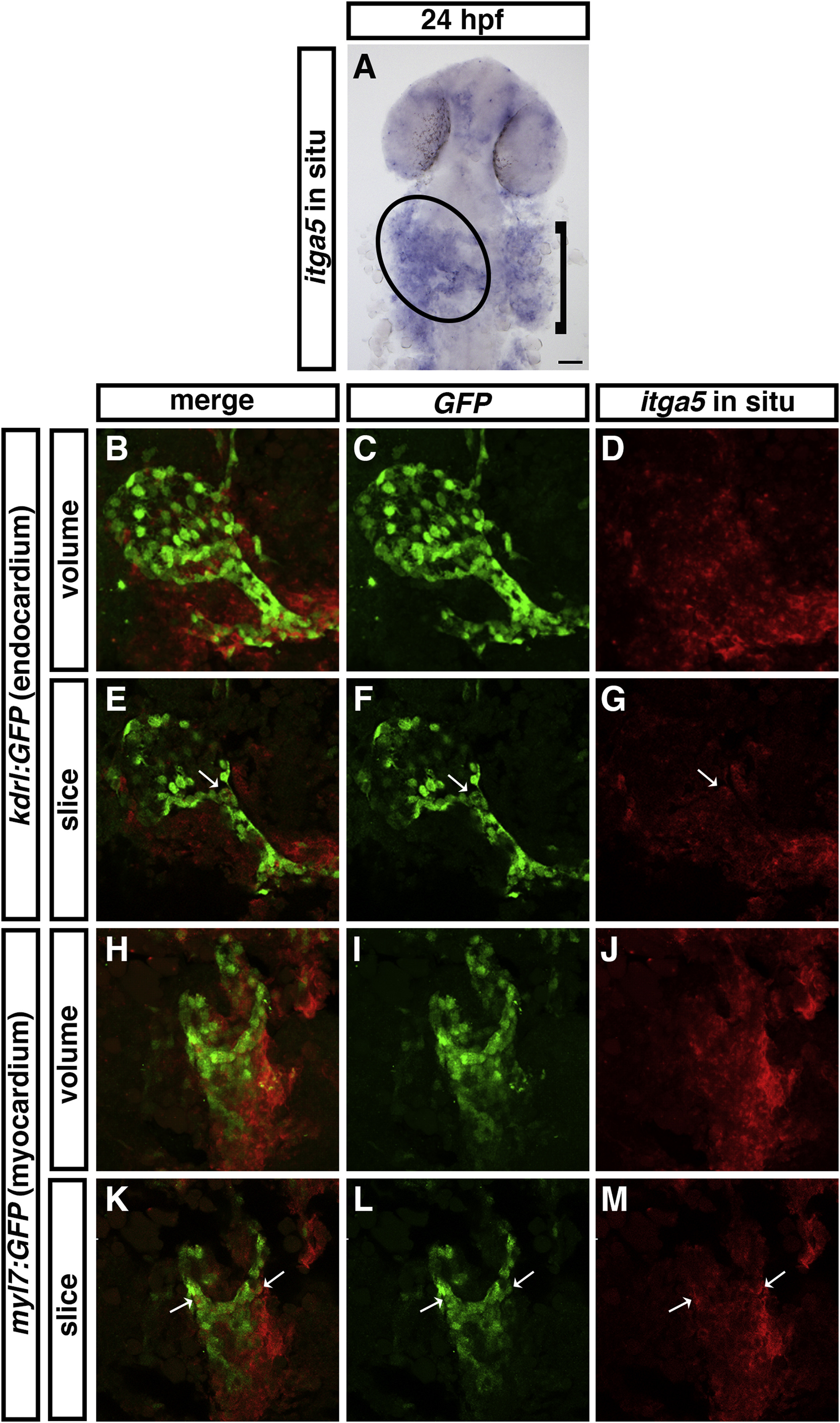
Fig. 1 Fig. 1. itga5 mRNA is expressed in the heart. (A) ISH for itga5 shows that it is enriched on the left side of the embryo at 24 hpf, indicated by the oval. Bracket indicates itga5 expression in the region of the developing pharyngeal arches. (B–D) Volume reconstructions of fluorescent ISH for itga5 (red, D) and immunofluorescence for kdrl:GFP (green, C) reveal that the itga5 expression domain broadly overlaps with the endocardial tube at 24 hpf. (E–G) A single slice from the Z-stack confirms colocalization of itga5 mRNA and GFP expression in an endocardial cell, indicated by white arrows. (H–J) Volume reconstructions of fluorescent ISH for itga5 (red, J) and immunofluorescence for myl7:GFP (green, I) reveal that the itga5 expression domain broadly overlaps with the myocardial tube at 24 hpf. (K–M) A single slice from the Z-stack confirms colocalization of itga5 mRNA and GFP expression in myocardial cells, indicated by white arrows. Dorsal views, anterior to the top.
Reprinted from Developmental Biology, 465(1), Schumacher, J.A., Wright, Z.A., Owen, M., Bredemeier, N.O., Sumanas, S., Integrin α5 and Integrin α4 cooperate to promote endocardial differentiation and heart morphogenesis, 46-57, Copyright (2020) with permission from Elsevier. Full text @ Dev. Biol.