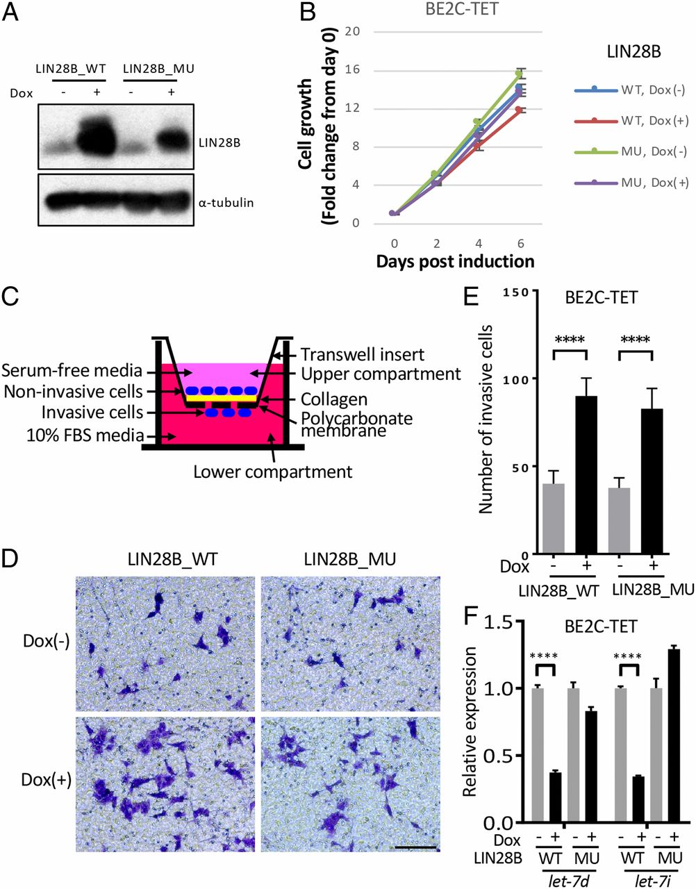
Fig. 3 Both WT and mutant LIN28B promote cell invasion and migration in BE2C cells. (A) Western blotting of LIN28B_WT/MU using the BE2C-TET cells untreated or treated with 50 ng/mL doxycycline for 3 d. α-Tubulin was used as a loading control. (B) Relative cell growth of BE2C-TET cells untreated or treated with 50-ng/mL doxycycline. Values represent means ± SEM of triplicate experiments. (C) Schematic of transwell migration and invasion assay. (D and E) Transwell migration and invasion assay of the BE2C-TET cells untreated or treated with 50-ng/mL doxycycline. Cells were stained with 0.1% crystal violet (D), and the number of migrated cells through the membrane per field was compared with the two-tailed unpaired t test (E). Values represent means ± SD of triplicate experiments. ****P < 0.0001. (Scale bar, 100 µm.) (F) qRT-PCR to detect let-7d and let-7i expressions in BE2C-TET cells that were either untreated or treated with 50-ng/mL doxycycline for 3 d. Values were normalized to U6, U47, and RNU44 small nuclear RNAs and represent the means ± SD of triplicate experiments. Statistical analysis was performed using the two-tailed unpaired t test. ****P < 0.0001.
Image
Figure Caption
Acknowledgments
This image is the copyrighted work of the attributed author or publisher, and
ZFIN has permission only to display this image to its users.
Additional permissions should be obtained from the applicable author or publisher of the image.
Full text @ Proc. Natl. Acad. Sci. USA

