IMAGE

Figure 3

ID
ZDB-IMAGE-200805-9
Source
Figures for Grivas et al., 2020
Image
Figure Caption

Figure 3

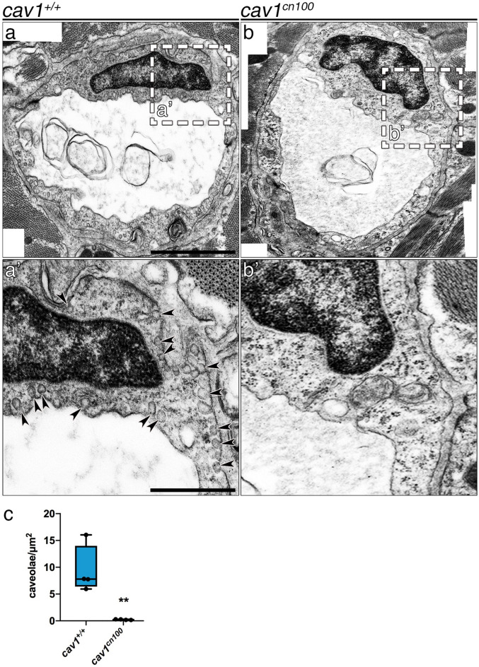
Loss of caveolae in cav1cn100 hearts. TEM images of cav1+/+ (a, a′) and cav1cn100 (b, b′) endothelial cells from the coronary vasculature. (a′, b′) Higher magnification of the dashed areas in (a) and (b). Arrowheads indicate membrane-bound caveolae. Scale bars: 1 μm in (a), (b); 0.5 μm in (a′), (b′). (c) Caveolae number per μm2 of endothelial cell. nWT = ncn100 = 4, t-test, **P < 0.01.

Acknowledgments
This image is the copyrighted work of the attributed author or publisher, and ZFIN has permission only to display this image to its users. Additional permissions should be obtained from the applicable author or publisher of the image. Full text @ Sci. Rep.