IMAGE

Fig. 4

ID
ZDB-IMAGE-200730-12
Source
Figures for Hall et al., 2020
Image
Figure Caption

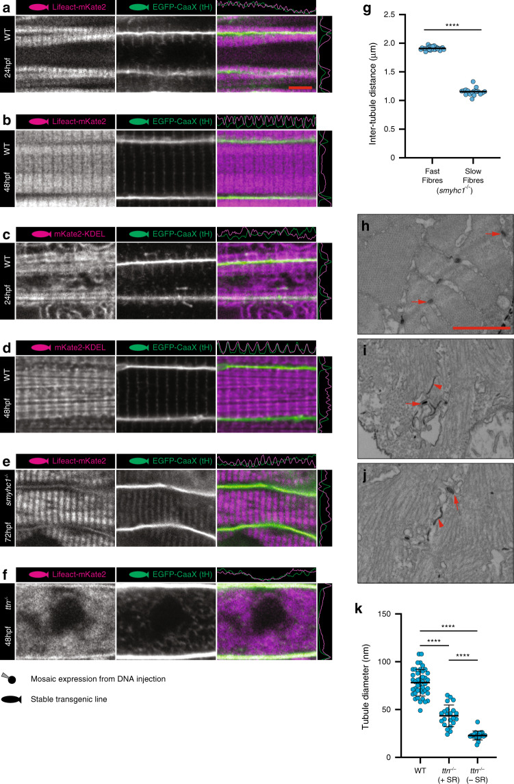
Fig. 4 Sarcomere formation and sarcoplasmic reticulum.

a, b Lifeact (marks actin). See also Supplementary Movie 4. c, d KDEL (marks endoplasmic/sarcoplasmic reticulum). esmyhc1/ embryos show intact tubules with reduced but regular spacing in slow muscle fibres. ftitin/ embryos show dysregulated tubule structure. g Quantification of inter T-tubule distance in fast and slow fibres from smyhc1/ mutant embryos. Fast fibres n = 25, slow fibres n = 17 biologically independent animals, one cell per animal over one independent experiment. Two-tailed T-test. Error bars show mean ± SD. **** p < 0.0001. h Transverse TEM section showing WT tubules (arrows). i, j Transverse TEM sections from titin/ mutants showing difference in morphology between tubules associated with sarcoplasmic reticulum (arrows) and tubules not associated with sarcoplasmic reticulum (arrowheads). k Quantification of sarcoplasmic reticulum associated and non-associated tubules in titin/ mutant embryos. WT n = 42, ttn/ +SR n = 22, ttn/ −SR n = 19 tubules from two biologically independent animals per group, over one independent experiment. One-way ANOVA followed by Tukey’s multiple comparison test. Error bars show mean ± SD. ****p < 0.0001. (a, c) 24 hpf; (b, d, f, k), 48 hpf (e g, h, i, j), 72 hpf. Scale bars (af) 5 µm, (hj) 1 µm. All images are representative of 12 individual cells within different individual animals.

Figure Data
Acknowledgments
This image is the copyrighted work of the attributed author or publisher, and ZFIN has permission only to display this image to its users. Additional permissions should be obtained from the applicable author or publisher of the image. Full text @ Nat. Commun.