
|
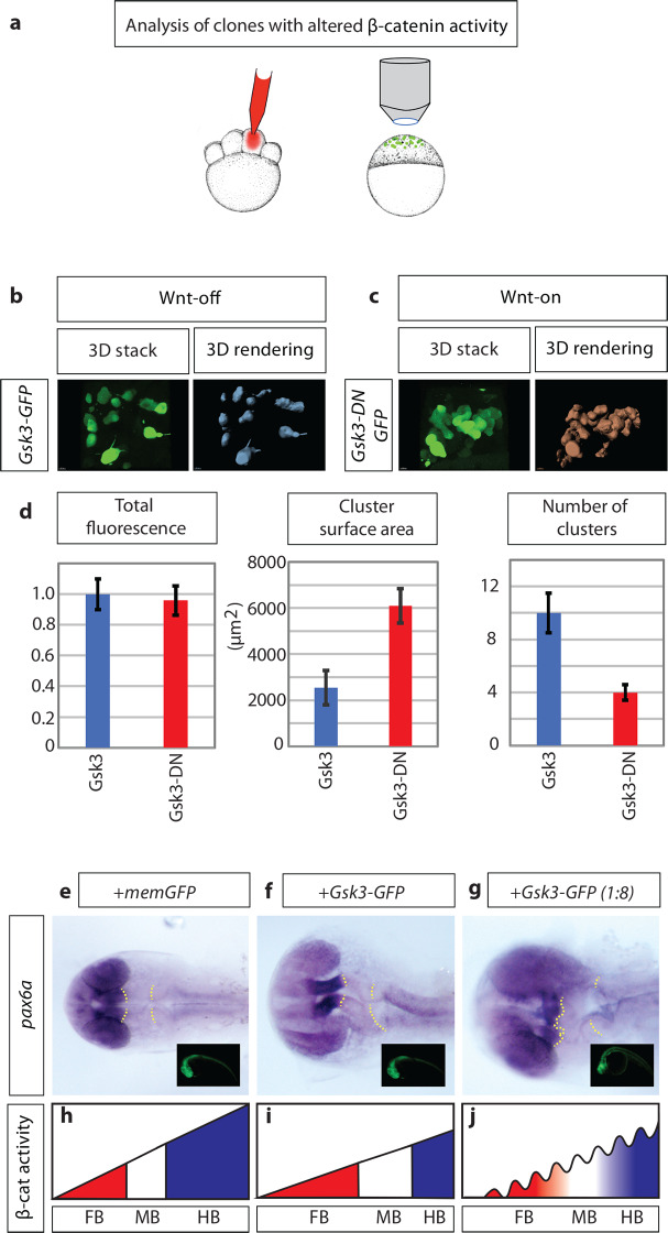
Fig 2
a) schematic illustration of the experimental procedure: 150ng mRNA injection of β-catenin effector Gsk3β into one cell at the eight blastomere stage. At sphere stage (prior induction of Wnt ligand expression), embryos were subjected to an image-based approach to analyse the distribution of cell clones in the animal tissue. b) expression of Gsk3β-GFP (Wnt-OFF) leads to a dispersed clone at the sphere stage. c) However, expression of dominant-negative Gsk3β-GFP (Gsk3β-DN-GFP; Wnt-ON) leads to clustering of the clonal cells. d) Gsk3β-DN expressing cells (Wnt-ON) show large clusters: demonstrated by an increased cluster surface and a reduced number of total clusters/cells compare to cells expressing WT Gsk3β (Wnt-OFF). The expression levels of Gsk3β and Gsk3β-DN are kept at a similar level shown by comparable total GFP-fluorescence in the clones. e)-g), embryos were injected with the indicated constructs and subjected to