
|
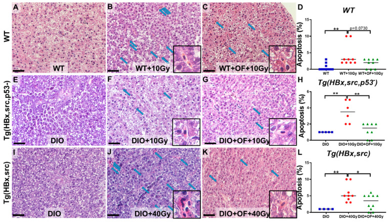
Figure 4
Representative images of hepatocyte apoptosis detected on hematoxylin and eosin (H&E) stain liver specimen from WT and transgenic fish with radiation without Oligo-Fucoidan or with Oligo-Fucoidan pretreatment. The images were taken at 400× magnification, and the scale shown is for 30 µm; the box area is enlarged to show the hepatocyte apoptosis, and the blue arrows pointed to the apoptotic hepatocytes. (