IMAGE

Figure 1

ID
ZDB-IMAGE-200713-20
Genes
Source
Figures for Coomer et al., 2020
Image
Figure Caption

Figure 1

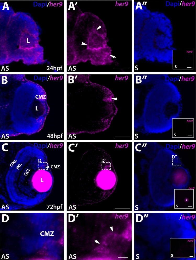
Her9 expression in the developing retina. Fluorescent in situ hybridization (FISH) showing her9 mRNA expression at 24 (AA′), 48 (BB′), and 72 (CC′) hpf in the developing retina (expression in the lens at 72 hpf is auto-fluorescence). (DD′) × 100 magnification of boxed area in (CC′) showing her9 expression in the CMZ. her9 sense probes at 24, 48, and 72 hpf are shown in (A″, B″, C″, D″). Panels in (A″D″) display her9 sense probe without the DAPI staining. L, lens CMZ, ciliary marginal zone; NR, neural retina; ONL, outer nuclear layer; INL, inner nuclear layer; GCL, ganglion cell layer. Scale bar = 50 μm and 100 μm.

Figure Data
Acknowledgments
This image is the copyrighted work of the attributed author or publisher, and ZFIN has permission only to display this image to its users. Additional permissions should be obtained from the applicable author or publisher of the image. Full text @ Sci. Rep.