IMAGE

FIGURE 1

ID
ZDB-IMAGE-200627-9
Genes
Source
Figures for Schredelseker et al., 2020
Image
Figure Caption

FIGURE 1

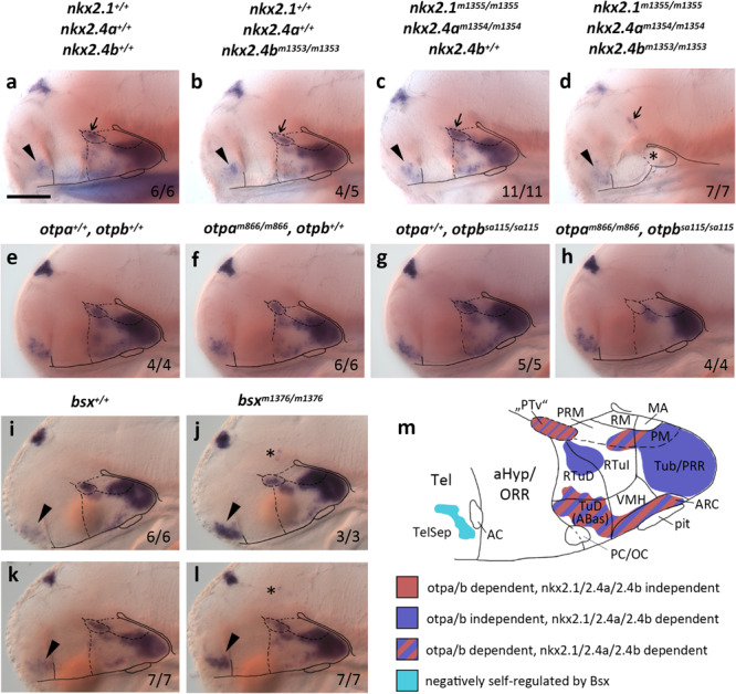
bsx expression depends on Nkx2.1, Nkx2.4, and Otp transcription factors. (a–l) Lateral views of wildtype and mutant (genotypes as indicated above panels) 4 dpf (a–d), 3 dpf (e–h,k,l) or 2 dpf (i,j) embryos, stained by in situ hybridization for bsx expression. All images are minimum intensity projections of 50 brightfield focal planes (distance 1 μm). Anterior is to the left. Scale bar (a–l): 100 μm. The ratio of observed phenotype and total analyzed embryos per genotype [n (phenotype)/n (total)] is indicated in right bottom corner of each panel. Arrowheads indicates bsx expression in the telencephalic septal region; small arrows indicate bsx expression which might correspond to “PTv” region; asterisk in (d) indicates tissue which might correspond to residual basal hypothalamus (bHyp) but which was not confirmed through marker gene expression in nkx2.1, nkx2.4a, nkx2.4b triple mutants; asterisks in (j,l) indicate ectopic bsx expressing cells. (m) Schematic drawing of a lateral view of bsx expressing forebrain regions of a zebrafish embryo 3–4 dpf illustrating which of the bsx expression domains depend on Otp or Nkx2.1 and Nkx2.4 transcription factors as indicated by color code. Anterior at left. For abbreviations see list.

Figure Data
Acknowledgments
This image is the copyrighted work of the attributed author or publisher, and ZFIN has permission only to display this image to its users. Additional permissions should be obtained from the applicable author or publisher of the image. Full text @ Front. Neurosci.