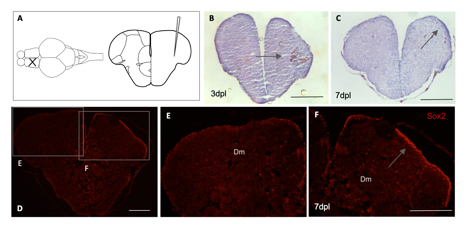
Fig. s3 Stab lesion and tissue integrity in adult zebrafish telencephalon. Schematic representation of lesion in the telencephalon area of adult zebrafish (A). Haematoxylin-Eosin staining and arrows indicate blood clot formation at 3 dpl (n = 3) (B) and apparent increase in cell number with higher intensity of staining at the lesioned hemisphere at 7dpl (n = 4) (C). Immunohistochemical labeling with Sox2 at 7 dpl indicates higher number of neural stem cells in the ventricular zone of the telencephalon (n = 3) (D). Magnification of image shown the contralateral hemisphere in E and regenerating hemisphere in F with increase in cells positive for Sox2 in the ventricular zone of the lesioned telencephalon. Scale bar (B-F): 200μM.
Image
Figure Caption
Acknowledgments
This image is the copyrighted work of the attributed author or publisher, and
ZFIN has permission only to display this image to its users.
Additional permissions should be obtained from the applicable author or publisher of the image.
Full text @ PLoS One

