
|
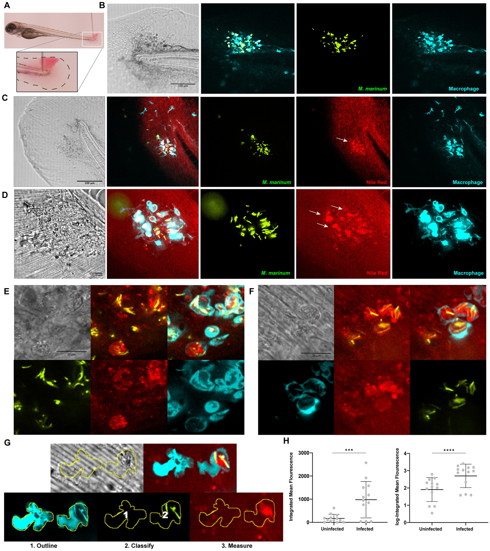
Fig. 2 Neutral lipid accumulation is observed during in vivo infections using vital staining. a. Larvae were injected at 3 dpf into the tail fin. A bacterial suspension was deposited between epithelial layers of the tail fin as shown. b. Confocal imaging of the tail fin of infected Tg(mfap4:p2A-Turquoise2) larvae at 2 days post infection (dpi) with M. marinum (green) showing macrophage (turquoise) recruitment to site of infection. Bar = 100 μm. c. Confocal imaging of Tg(mfap4:p2A-Turquoise2) larvae 2 dpi with M. marinum (green) and following staining with Nile red. Arrows indicate location of neutral lipid accumulation within infected macrophages. Bar = 100 μm. d. Confocal imaging of Tg(mfap4:p2A-Turquoise2) larvae 2 dpi with M. marinum (green) and following staining with Nile red. Arrows indicate location of neutral lipid accumulation within infected macrophages. 60x objective, bar = 10 μm. e. Confocal imaging of a representative Tg(mfap4:p2A-Turquoise2) larva 2 dpi with M. marinum (green) and following staining with Nile red showing detail of Nile red staining pattern. 60x objective, bar = 20 μm. f. Confocal imaging of a representative Tg(mfap4:p2A-Turquoise2) larva 2 dpi with M. marinum (green) followed by staining with Nile red showing detail of Nile red staining pattern. 60x objective, scale bar = 20 μm. g. Representative images of uninfected and infected macrophages depicting the method of quantification of Nile red fluorescence intensity by first outlining all macrophages, then classifying uninfected (1) vs. infected (2) macrophages, and then measuring the integrated mean Nile red fluorescence of uninfected vs. infected macrophages. h. Integrated mean fluorescence and log-Integrated mean fluorescence of Nile red in uninfected vs. infected macrophages. Each data point represents the mean value of all macrophages within each class for a given animal. Data were collected from 13 individual animals. ***p = 0.0002 (Wilcoxon matched-paired sign rank test), ****p<0.0001 (paired t-test).