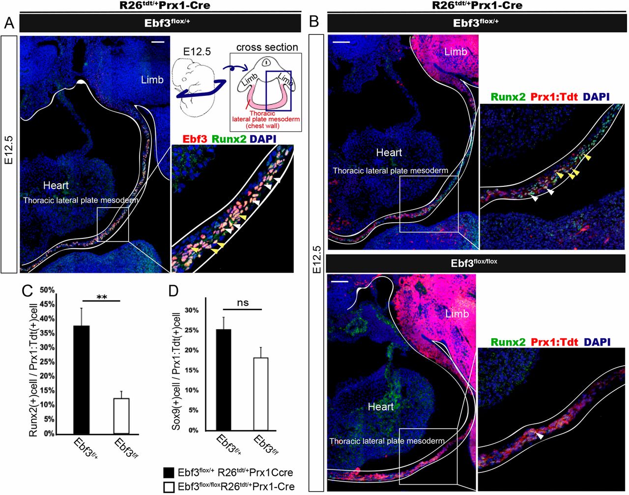
Fig. 4 Decreased number of Runx2+ pre-osteoblasts in Ebf3-deficient thoracic lateral plate mesoderm at E12.5. (A) Transverse section of thoracic lateral plate mesoderm (as indicated in the schematic) at E12.5 immunostained with anti-Ebf3 and anti-Runx2 antibodies. Most but not all Ebf3-expressing cells were Runx2 positive. White and yellow arrowheads show Ebf3/Runx2 double-positive cells. (B) Decrease in the number of Prx1: Tdtomato+ Runx2+ cells in Ebf3flox/flox Prx1-Cre compared with that in heterozygous embryos (white arrowheads). Immunostained transverse sections of the thoracic lateral plate mesoderm at E12.5 are shown. A and upper panels of B show images of Ebf3/Runx2-double-positive cells and those of Prx1: Tdtomato+Runx2+ LPMs, respectively, prepared from a single section of a heterozygous embryo stained with antibodies against Ebf3, Runx2 and DAPI. Yellow arrowheads indicate Prx1: Tdtomato+Ebf3+Runx2+ LPMs. (C) The ratio of Runx2+ cells in Prx1: Tdtomato+ thoracic LPMs was lower in Ebf3flox/flox Prx1-Cre KO mice than in Ebf3 heterozygous mice at E12.5. Error bars represent s.e.m. **P<0.01 (n=4). (D) Ratio of Sox9+ cells in Prx1: Tdtomato+ thoracic LPMs at E12.5. Error bars represent s.e.m.; ns, non-significant (n=4). Scale bars: 100 μm
Image
Figure Caption
Acknowledgments
This image is the copyrighted work of the attributed author or publisher, and
ZFIN has permission only to display this image to its users.
Additional permissions should be obtained from the applicable author or publisher of the image.
Full text @ Development

