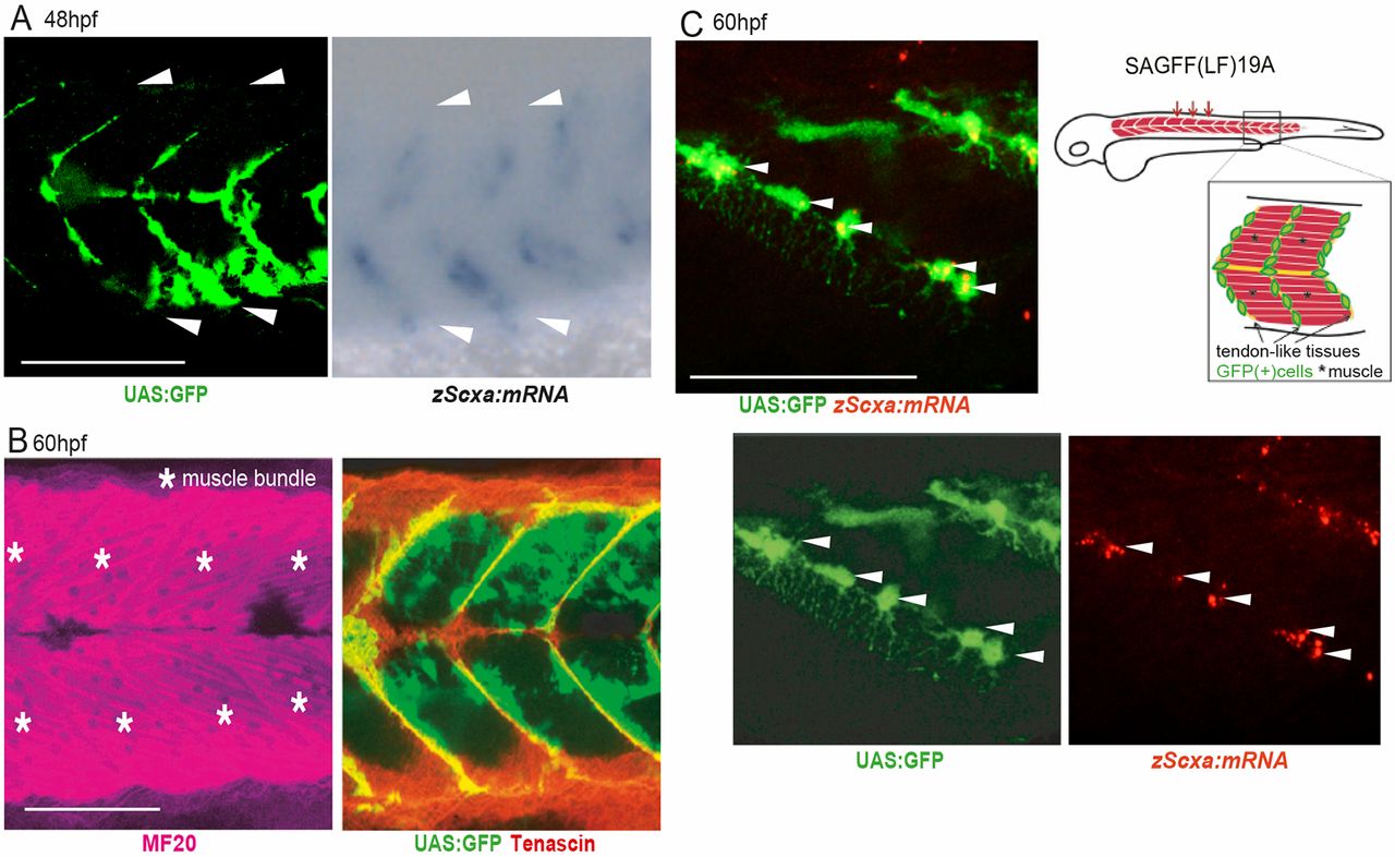
Fig. 1 Identification of an enhancer trap line marking progenitors of tenocytes and interstitial cells in zebrafish embryos. (A) Expression of UAS:GFP in Gal4-UAS enhancer trap zebrafish line SAGFF(LF)19A and signals for scxa mRNA transcripts detected by whole-mount in situ hybridization (WISH) at 48 hpf. White arrowheads show segmental boundaries of somites. (B) Whole-mount immunostaining of 60 hpf SAGFF(LF)19A embryos for myogenic (magenta, MF20) and tenogenic (red, tenascin) markers together with UAS:GFP signals at 60 hpf. Asterisks mark muscle bundles. (C) Tenocyte progenitors marked with UAS:GFP and fluorescence in situ hybridization for scxa (red) at 60 hpf. Note that these cells extended long filopodia through the boundaries of skeletal muscle fibers. Arrowheads indicate UAS:GFP+ zScxa:mRNA+ cells. Schematic shows UAS:GFP+ cells in tendon and connective tissues in trunk muscles of SAGFF(LF) 19A line. Scale bars: 100 μm (A,B); 50 μm (C).
Image
Figure Caption
Acknowledgments
This image is the copyrighted work of the attributed author or publisher, and
ZFIN has permission only to display this image to its users.
Additional permissions should be obtained from the applicable author or publisher of the image.
Full text @ Development

