
|
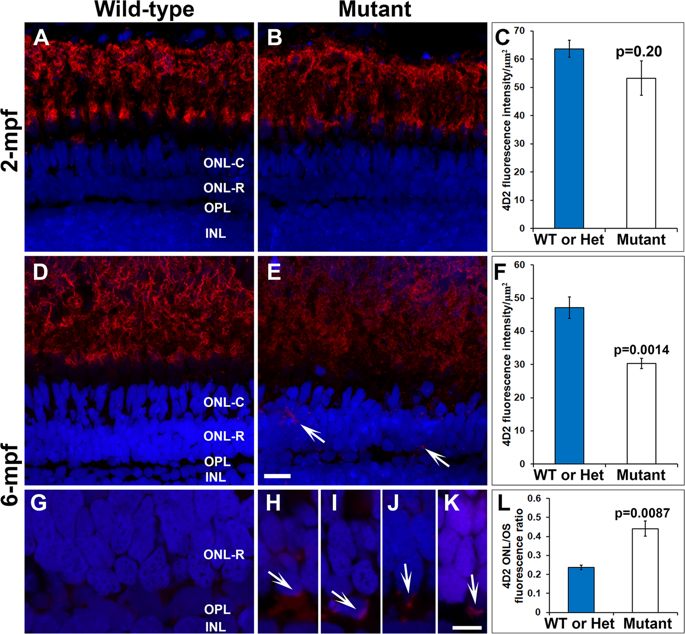
Fig. 6
Reduction and mislocalization of rhodopsin in pomgnt1 mutant retina. Retinal sections from 2-mpf and 6-mpf zebrafish were immunostained with antibody 4D2 (red) and counter-stained with DAPI (blue). (A) 4D2 immunostaining of wild-type retina at 2-mpf. (B) 4D2 immunostaining of homozygous pomgnt1sny7 mutant retina at 2-mpf. (C) Quantification of 4D2 fluorescence intensity at 2-mpf. There was no significant reduction in 4D2 intensity in the mutant fish at this age. Student’s t-test. (D) 4D2 immunostaining of wild-type retina at 6-mpf. (E) 4D2 immunostaining of homozygous pomgnt1sny7 mutant retina at 6-mpf. Note the reduction in 4D2 fluorescence intensity. Note the mislocalization of 4D2 immunoreactivity to the ONL-R (arrows). (F) Quantification of 4D2 fluorescence intensity at 6-mpf. There was a significant reduction in 4D2 intensity in the mutant fish at this age. Student’s t-test. (G) High mag of wild-type 4D2 staining at 6-mpf. Fluorescence intensity of 4D2 in the ONL-R was low. (H–K) High mag of homozygous pomgnt1sny7 mutant 4D2 staining at 6-mpf. Note the mislocalization of 4D2 immunoreactivity to the ONL-R layer (arrows). (L) Ratio of 4D2 fluorescence intensity of outer nuclear layer/outer segment layer (ONL/OS). 4D2 ONL/OS fluorescence ratio was increased in the mutants. Student’s t-test. Scale bar in E: 5 µm for A, B, D and E; scale bar in K: 5 µm for G-K.