
|
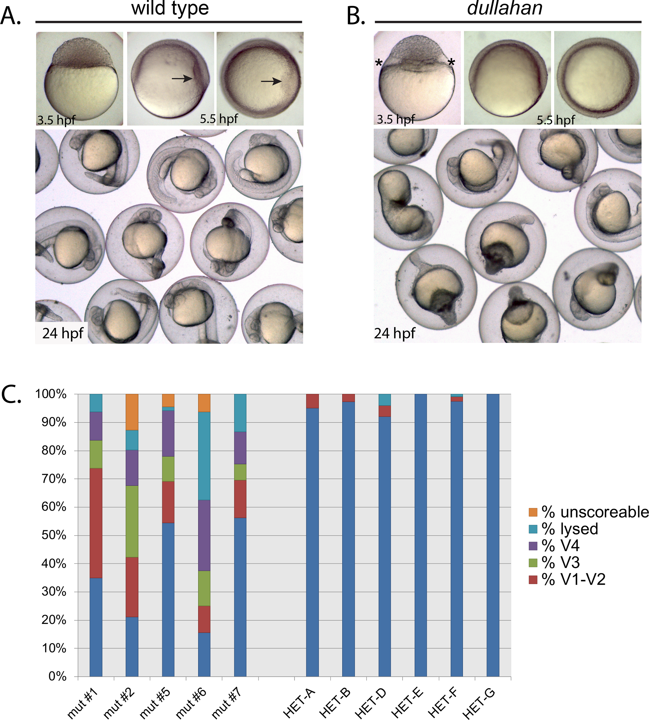
Fig. 5 dullahan mutant phenotype. A. Wild-type and (B) dul embryos at 3.5, 5.5, and 24 hpf. Embryos in upper left and center panels are lateral views, and upper right panels are animal pole views. The enlarged cytoplasmic region between the yolk and blastomeres in the dullahan mutant at 3.5 hpf is noted with asterisks. The dorsal shield (arrow) is to the right in the 5.5 hpf wild-type embryo and absent in the mutant. C. Distribution of embryonic phenotypes from five dul females (left) compared to 6 siblings (right): mut #1 (n = 80), mut #2 (n = 71), mut #5 (n = 68), mut #6 (n = 32), mut #7 (n = 105), HET-A (n = 103), HET-B (n = 112), HET-C (n = 76), HET-D (n = 76), HET-A (n = 67), HET-A (n = 118), HET-G (n = 32). In each cross heterozygous or mutant females were crossed to wild-type (TL) males.