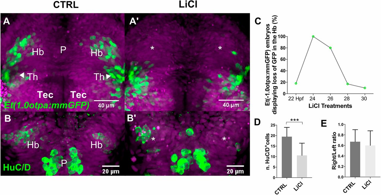
Fig. 2 Premature intrinsic activation of Wnt signaling delays habenular neuron differentiation. (A-B′) Projections of confocal z-stacks, dorsal views, anterior is towards the top focused onto the diencephalon of (A,A′) Et(-1.0otpa:mmGFP) and (B,B′) tg(flh:GFP); tg(foxD3:GFP) transgenic embryos. Nuclei are DAPI labeled (purple). (A,A′,C) Transient Wnt signaling activation causes a specific loss of GFP-expressing habenular neurons at 48 hpf. (B,B′,D,E) The number of HuC/D-positive differentiating habenular neurons is reduced at 36 hpf; their left-right asymmetric development remains unchanged. CTRL, control; Hb, habenulae; P, pineal; Tec, optic tectum; Th, thalamus.
Image
Figure Caption
Figure Data
Acknowledgments
This image is the copyrighted work of the attributed author or publisher, and
ZFIN has permission only to display this image to its users.
Additional permissions should be obtained from the applicable author or publisher of the image.
Full text @ Development

