
|
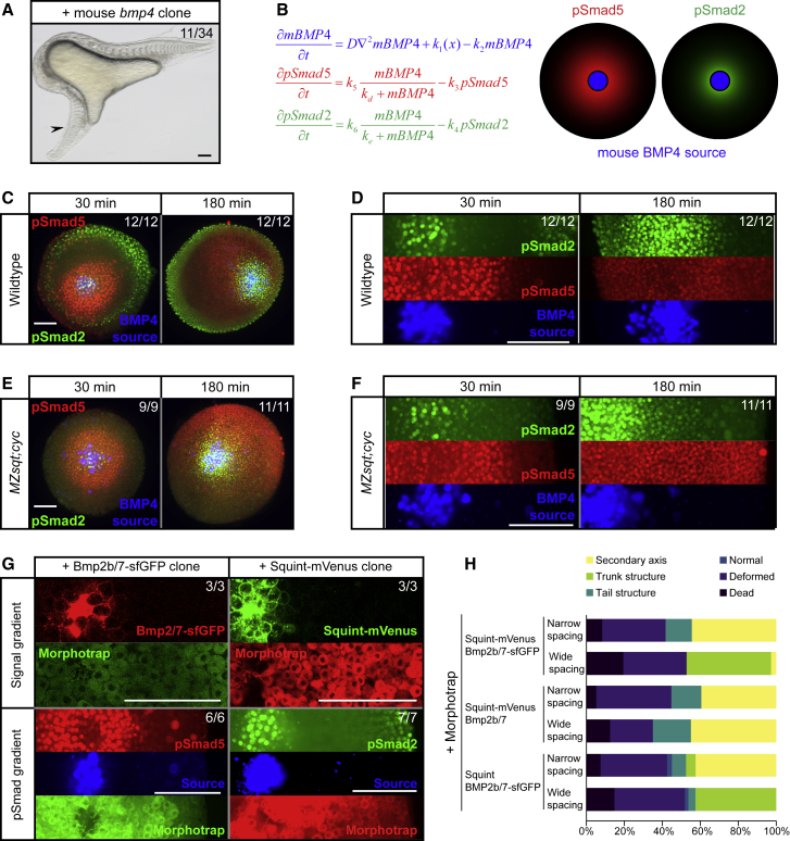
Figure 3
Different BMP and Nodal Signaling Ranges Arise from Differential Signaling Activation Kinetics
(A) Single clones expressing mouse
(B) Mathematical modeling shows that a difference in signaling activation kinetics could explain how a single gradient of mBMP4 induces pSmad5 and pSmad2 at different ranges.
(C) Wild-type zebrafish embryos with clones expressing
(D) Higher magnification of images shown in (C) with separated fluorescent channels. Scale bar, 150 μm.
(E)
(F) Higher magnification of images shown in (E) with separated fluorescent channels. Scale bar, 150 μm.
(G) Zebrafish Bmp2b/7-sfGFP and Squint-mVenus clones in morphotrap-expressing wild-type embryos 30 min post-transplantation. Scale bar, 150 μm.
(H) Double clones with fluorescently tagged or untagged zebrafish Nodal and BMP and with narrow or wide spacing were generated in morphotrap-expressing embryos. The frequency of the different structures induced by the clones was assessed 24 h post-transplantation.