
|
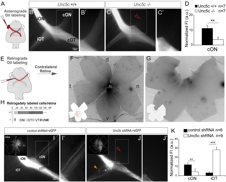
Figure 4
Unc5c Is Necessary for Establishment of the Retino-retinal Projection
(A–C) DiI was placed into one eye and the labeled axons viewed at the chiasm (A).
(B and C) DiI-labeled E17.5 Unc5c+/+ and Unc5c−/− embryos. The fluorescence intensity in the contralateral optic nerve of the Unc5c−/− embryo is decreased compared with the control littermate (arrowhead).
(B’ and C’) Higher magnification of the boxed regions in (B) and (C).
(D) Mean (± SEM) normalized fluorescence intensity (FI) in the contralateral optic nerve of E17.5 Unc5c−/− and wild-type embryos monocularly injected with DiI.
(E–G) DiI was applied to one optic nerve and the opposite retina analyzed (E).
(F and G) Whole-mounted retinas from E17.5 Unc5c+/+ and Unc5c−/− embryos retrogradely labeled with DiI. Inserts: tracings of the labeled cells are shown.
(H) Mean (± SEM) number of retrogradely labeled cells in each quadrant of E17.5 wild-type and Unc5c mutant retinas.
(I and J) E16.5 embryos electroporated at E13.5 with control or Unc5c shRNAs plus EGFP-encoding plasmids. Insert: corresponding whole-mounted electroporated retina is shown. EGFP-positive axons were present in the contralateral optic nerve of control (I and I’), but not Unc5c shRNA electroporated, embryos (open arrowhead, J and J’). Unc5c electroporated embryos also displayed an ectopic ipsilateral projection (orange arrowhead).
(K) Mean (± SEM) normalized fluorescence intensity in the contralateral optic nerve and ipsilateral optic tract of E16.5 embryos electroporated at E13.5 with Unc5c shRNA or control shRNA.
cON, contralateral optic nerve c; cOT, contralateral optic tract; D, dorsal; iON, ipsilateral optic nerve; iOT, ipsilateral optic tract; N, nasal; T, temporal; V, ventral. Error bars indicate ± SEM. (∗∗p < 0.01, ∗∗∗p < 0.001, Student’s unpaired t test).
See also