
|
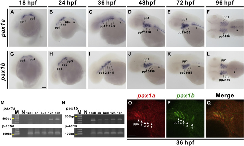
Fig. 1 Developmental expression pattern of pax1a and pax1b. Signals for pax1a (A–F) and pax1b (G–L) were observed in the developing pharyngeal pouches (pp) and sclerotome (s) from 18 to 96 hpf. pax1a- or pax1b-hybridized embryos are shown from a lateral view at 18 hpf (A, G), 24 hpf (B, H), 36 hpf (C, I), 48 hpf (D, J) and 72 hpf (E, K), and a dorsal view is shown for 96 hpf (F, L). Semi-quantitative RT-PCR indicates expression of pax1a (M) and pax1b (N) from 1 cell to 18 hpf stages. Expression of β-actin was used as a control. Molecular weight marker (M) and no template control (N) are also shown. Double fluorescence in situ hybridization revealed co-expression of pax1a (O) and pax1b (P) in pharyngeal pouches 1–5 at 36 hpf. Merged image (Q) is shown. Lateral view of embryos is shown with anterior aspect to the right. Sh, shield. Scale bars represent 100 μm.
Reprinted from Mechanisms of Development, 161, Liu, Y.H., Lin, T.C., Hwang, S.L., Zebrafish Pax1a and Pax1b are required for pharyngeal pouch morphogenesis and ceratobranchial cartilage development, 103598, Copyright (2020) with permission from Elsevier. Full text @ Mech. Dev.