
Image
|
Figure Caption
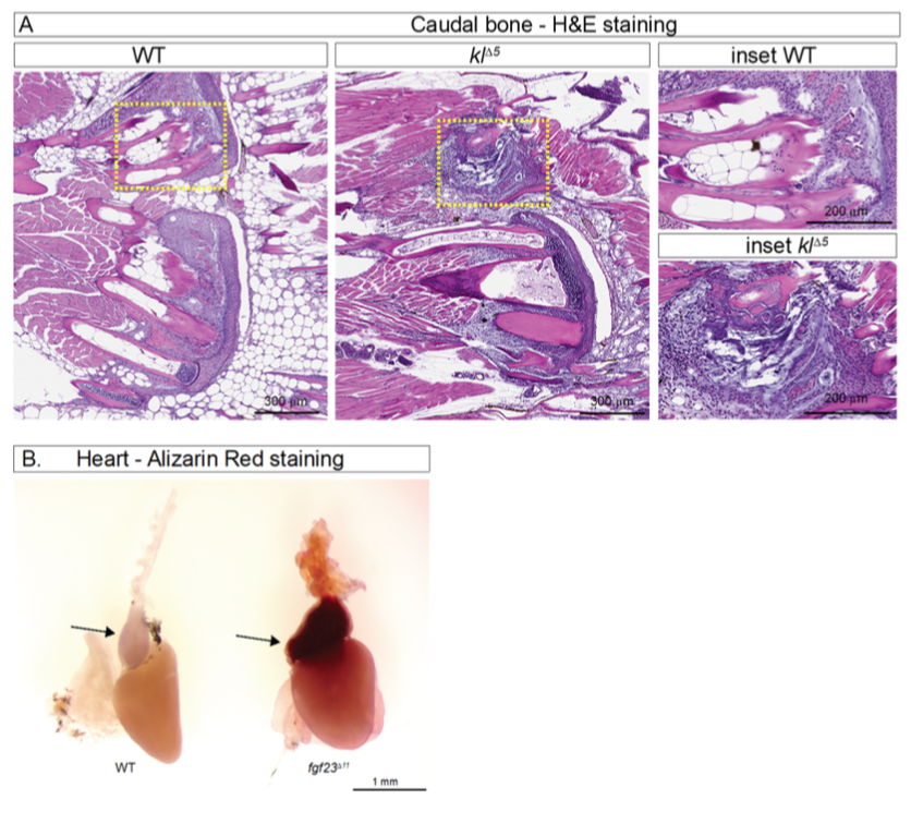
Fig. S2
Histological characterization of zebrafish heart. (A) H&E-stained serial sections across caudal bone of wildtype and αklotho mutant; dashed yellow lines demarcate region enlarged in inset. Genotypes as indicated in the image (n=3 males, 5 months post-fertilization). (B) Alizarin red staining on fgf23 mutant hearts to visualize calcification. Alizarin red staining on whole-mount hearts of background control (AB; left) and fgf23 mutant (fgf23Δ11; right). Intense red staining is observed in the outflow tract of the heart (arrow) in fgf23 mutant. (n=5 males, 5 months post-fertilization).
Acknowledgments
This image is the copyrighted work of the attributed author or publisher, and
ZFIN has permission only to display this image to its users.
Additional permissions should be obtained from the applicable author or publisher of the image.
Full text @ Cell Rep.