IMAGE

Fig. 5

ID
ZDB-IMAGE-200201-16
Antibodies
Source
Figures for Iribarne et al., 2019
Image
Figure Caption

Fig. 5

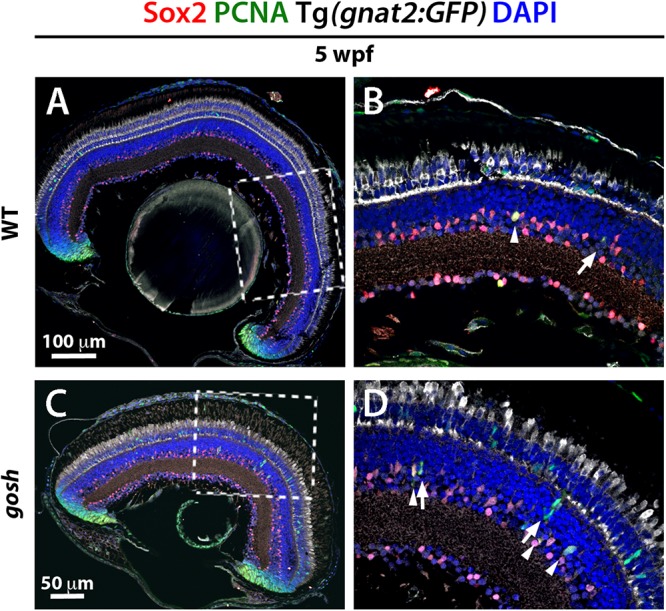
Müller glia express Sox2 during the regeneration response in gosh mutants. Wild-type and gosh mutant retinas at 5 wpf labeled with anti-Sox2 and anti-PCNA antibodies, and DAPI. Tg(gnat2:GFP) transgene visualizes cones. Wild-type retinas display Sox2 expression in the CMZ and amacrine cells (A,C, dotted boxes show the area magnifies in B,D). Müller glia exhibit faint expression of Sox2. At 5 wpf, gosh mutant retinas show that Sox2 expression is colocalized with PCNA in the INL (D, arrowheads) probably in Müller glia, whereas subsequently differentiating NPCs express only PCNA (D, arrows).

Figure Data
Acknowledgments
This image is the copyrighted work of the attributed author or publisher, and ZFIN has permission only to display this image to its users. Additional permissions should be obtained from the applicable author or publisher of the image. Full text @ Front Cell Dev Biol