IMAGE

Figure 2

ID
ZDB-IMAGE-191230-1501
Source
Figures for Collodet et al., 2019
Image
Figure Caption

Figure 2

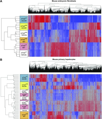
Hierarchical clustering of the transcriptome data. Overview of the hierarchical clustering analyses in MEFs (A) and mouse primary hepatocytes (B). Mean-centered gene expression ratios are depicted by a log2 pseudocolor scale; red indicates that the gene is overexpressed compared to the mean value, whereas blue specifies that the gene is less expressed. Data were analyzed by 2-way ANOVA with the factors of genetic background, treatment, and interaction. Only selected genes having a FDR of P < 0.0001 for the interaction are shown.

Acknowledgments
This image is the copyrighted work of the attributed author or publisher, and ZFIN has permission only to display this image to its users. Additional permissions should be obtained from the applicable author or publisher of the image. Full text @ FASEB J.