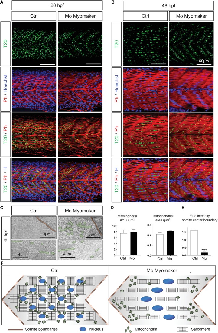
Mitochondrial network maturation is conditional to myoblast fusion. (A,B) Confocal imaging of mitochondrial network (Tomm20, T20; green) counterstained with Phalloidin (Ph; red) and Hoescht (H; blue) at 28 hpf (A) and 48 hpf (B); control (Ctrl, mock injection) or Myomaker-targeting Morpholinos (Mo Myomaker). (C) Electron micrographs of longitudinal sections at 48 hpf in control and Myomaker depleted embryos. Top images are close to boundaries, bottom images are at the somite center. (D) Quantification of mitochondrial number and area at 48 hpf from longitudinal electron micrographs (n = 6 micrographs for Ctrl, n = 7 micrographs for Mo). (E) Quantification of Tomm20-zsGreen fluorescence ratio between somite center and boundary region 48 hpf (n = 4 fish per group, 3 images analyzed per fish). (F) Cartoon representing somite structure and mitochondrial network at 48 hpf in control and Myomaker depletion. Bars are mean ± SEM. ∗∗∗P < 0.001, unpaired T-test.
This image is the copyrighted work of the attributed author or publisher, and
ZFIN has permission only to display this image to its users.
Additional permissions should be obtained from the applicable author or publisher of the image.
Full text @ Front Cell Dev Biol
Your Input Welcome
Thank you for submitting comments. Your input has been emailed to ZFIN curators who may contact you if
additional information is required.
Oops. Something went wrong. Please try again later.