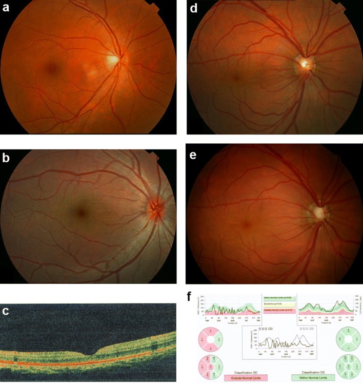
Fundus changes associated with high hyperopia in family ZOC710536. a Normal fundus of an unrelated normal control. b Fundus photograph of V:1. The right eye for the affected individual V:1 in Fig. 1 at 6 years old with refraction of + 12D/OS and + 12D/OS, and an axial length of 17.49 mm/OD and 17.53 mm/OS. Fundus change is typical for high hyperopia with a relatively normal fovea. c OCT scan of V:1. The right eye for the affected individual V:1 in Fig. 1 at 6 years old with refraction of + 12D/OS and + 12D/OS and an axial length of 17.49 mm/OD and 17.53 mm/OS. d Fundus photos of IV: 7 at 29 years old. The right eye for the affected individual IV:7 in Fig. 1 at 29 years old, with refraction of + 10.00 DS/OD and + 10.00 DS/OS and an axial length of 17.6 mm/OD and 17.47 mm/OS. e Fundus photos of IV: 7 at 38 years old. The right eye for the individual IV:7 in Fig. 1 at 38 years old, 2 weeks after the onset of angle-closure glaucoma in the right eye (with IOP 43 mmHg before treatment). An enlarged optic disc was observed compared with the fundus photograph in D. f Heidelberg retina tomograph (HRT) results for the affected individual IV:7 from Fig. 1. The HRT result in F demonstrated partial loss of the retinal ganglion cell layer in the temporal half of the retina in the right eye. OD right eye, OS left eye
Acknowledgments
This image is the copyrighted work of the attributed author or publisher, and
ZFIN has permission only to display this image to its users.
Additional permissions should be obtained from the applicable author or publisher of the image.
Full text @ Hum. Genet.
Your Input Welcome
Thank you for submitting comments. Your input has been emailed to ZFIN curators who may contact you if
additional information is required.
Oops. Something went wrong. Please try again later.